
Reflected Light Digital Image Gallery
Jumping Spider (Phidippus species)
As members of the Family Salticidae (jumping spiders), species in the genus Phidippus feature eight eyes, two body segments, and spinnerets. Some of the largest salticids in the world are found in Florida, such as P. regius and P. otiosus, and at least 12 species occur in the Sunshine State, from a genus that includes more than 51 described species. Worldwide in distribution, jumping spiders are particularly well studied and appreciated in Japan.
View a second image of a jumping spider (Phidippus species).
View a third image of a jumping spider (Phidippus species).
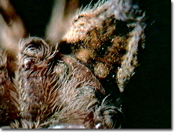
The eyes of a jumping spider are arranged with four on top of the head, or cephalothorax, and four in front. Phidippus spiders have excellent vision with some of the highest acuities in the invertebrate world. The larger anterior median eyes of the spider function similarly to the human fovea, with a narrow field of view, but very high acuity. The remaining six eyes provide excellent peripheral vision to these carnivorous arachnids, with a much broader field of view, but lower resolution. Featuring a long focal length, the anterior median eyes are long and tubular, which improves resolution and magnification. Because of the narrow field of vision exhibited by these high-resolution eyes, the spider takes in its visual cues by moving its head, and the eyes themselves, to scan for potential prey or predators. The motion of the eyes, however, is restricted to movement of the retinas rather than the eyeballs, because the lenses are fixed to the chitinous carapace. One can actually look into the eyes of a jumping spider and observe its eyes changing colors. The darkest appearance of the eyes is seen when the spider is looking straight ahead, because one is looking directly into the retina.
Jumping spiders utilize a relatively large proportion of their brains for visual processing. It is interesting to note that the proportion of brain-to-body devoted by a jumping spider to visual tasks is similar to a human's. These arachnids are commonly called jumping spiders because they are capable of jumping 25 times their body length, and as predators, accurately pounce on their prey instead of spinning webs for entrapment.
Adult females have small palpi that they use solely as tiny "hands" to manage prey and clean their faces. Adult male Phidippus spiders feature swollen palpi that are structurally more complex from the female apparatus, and are used to transfer sperm to the females. Unusual in the animal kingdom, there is an additional sperm duct of the male that opens up into the facial palp. These characteristics are used by arachnologists to differentiate the sexes of mature salticid spiders.
When the male is ripe, he spins a small web and deposits on it a drop of sperm from the underside of his abdominal region. The tip of the palp is placed in the sperm web and the sperm is drawn through the opening and into the palp sperm duct for storage and later transfer. Once the male locates a female, and if she is receptive to his courtship ritual dance, the male places his palp against an abdominal hard plate known as her epigynum, locks into place with his tibial apophysis placed in a groove at the posterior of her epigynum, and then the palpus expands. With increasing blood pressure, the sperm is injected into the female, transferred into hardened ducts and reservoirs, and stored until her eggs are ready to be fertilized and laid.
Contributing Authors
Cynthia D. Kelly, Thomas J. Fellers and Michael W. Davidson - National High Magnetic Field Laboratory, 1800 East Paul Dirac Dr., The Florida State University, Tallahassee, Florida, 32310.
BACK TO THE REFLECTED LIGHT IMAGE GALLERY
BACK TO THE DIGITAL IMAGE GALLERIES
Questions or comments? Send us an email.
© 1995-2025 by Michael W. Davidson and The Florida State University. All Rights Reserved. No images, graphics, software, scripts, or applets may be reproduced or used in any manner without permission from the copyright holders. Use of this website means you agree to all of the Legal Terms and Conditions set forth by the owners.
This website is maintained by our
Graphics & Web Programming Team
in collaboration with Optical Microscopy at the
National High Magnetic Field Laboratory.
Last Modification Friday, Nov 13, 2015 at 02:19 PM
Access Count Since September 17, 2002: 12800
Visit the website of our partner in introductory microscopy education:
|
|