
Polarized Light Digital Image Gallery
Uridine
As one of the four nucleosides used in genetic coding for RNA, uridine contains a pyrimidine base (uracil), which is structurally related to thymidine (linked to a ribose sugar) found in DNA strands. In RNA, the complement to uridine is the nucleoside adenosine and when a phosphate is attached, the nucleotide uridine monophosphate (UMP) is formed.
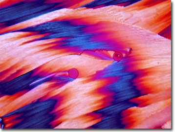
With 9 carbons, 12 hydrogens, 2 nitrogens, and 6 oxygen atoms per molecule, uridine is known to organic chemists as 1-beta-D-ribofuranosyluracil and features a molecular weight of 244.20. Known also as a riboside, uridine is extracted from yeast ribonucleic acids using a weak alkali solution. Crystalline needles of purified uridine melt at 165 degrees Celsius and are soluble in water. Nucleotides such as uridine monophosphate exist in their di- (UDP) and tri-phosphorylated (UTP) forms in the cells of an organism.
Many nucleotide analogues, including 5-fluorouracil and 5-iodo-2'-deoxyuridine, are chemically synthesized for their therapeutic abilities. As antiviral agents, nucleotide analogues interfere with replication of DNA/RNA and protein synthesis on ribosomes. When used in organ transplants, some uridine derivatives reduce the likelihood of rejection by suppressing the immune system of the host. Mitochondrial disease, characterized by problems with neurological, cognitive, kidney, and muscular functions, shows some improvement with treatment with triacetyluridine (TAU). A precursor, TAU rapidly converts to uridine, when taken orally. The mitochondria, essential for many cellular functions including energy production, are the sites of biosynthesis of uridine. A dietary supplement of uridine seems to overcome symptoms associated with this relatively rare genetic disease.
Contributing Authors
Omar Alvarado, Thomas J. Fellers and Michael W. Davidson - National High Magnetic Field Laboratory, 1800 East Paul Dirac Dr., The Florida State University, Tallahassee, Florida, 32310.
BACK TO THE POLARIZED LIGHT IMAGE GALLERY
BACK TO THE DIGITAL IMAGE GALLERIES
Questions or comments? Send us an email.
© 1995-2025 by Michael W. Davidson and The Florida State University. All Rights Reserved. No images, graphics, software, scripts, or applets may be reproduced or used in any manner without permission from the copyright holders. Use of this website means you agree to all of the Legal Terms and Conditions set forth by the owners.
This website is maintained by our
Graphics & Web Programming Team
in collaboration with Optical Microscopy at the
National High Magnetic Field Laboratory.
Last Modification Friday, Nov 13, 2015 at 02:19 PM
Access Count Since September 17, 2002: 12691
Visit the website of our partner in introductory microscopy education:
|
|