
Polarized Light Digital Image Gallery
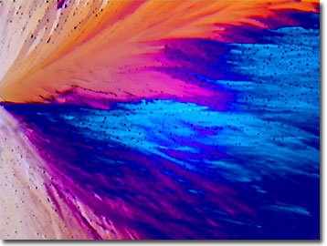
Erythromycin
Erythromycin is a wide-spectrum antibiotic that is used in ophthalmic, topical, and systemic treatment of bacterial infections. It is particularly useful in patients with a history of allergies towards other antibiotics, such as penicillin.
View a second image of erythromycin.
Naturally occurring, erythromycin is produced by a strain of the mold Streptomyces erythraeus and is classified as a macrolide antibiotic, as is clarithromycin and azithromycin. Erythromycin is basic and readily forms salts with acids, but it is the base form that is microbiologically active. Erythromycin base forms white to off-white crystals or powder that is slightly soluble in water, alcohol, and in organic solvents such as chloroform and ether. With normal hepatic functions, erythromycin is concentrated in the liver and excreted in the bile.
The mechanism of action appears to be binding to the 50S ribosomal subunits in susceptible bacteria to suppress protein synthesis, but the antibiotic does not affect nucleic acid synthesis. Delayed-release tablets of erythromycin are specially enteric-coated to protect the active antibiotic ingredients from the inactivating effects of gastric acidity and to permit efficient absorption in the small intestine. Side effects include fever, nausea, skin rash, pain, and swelling, and more rarely, allergic reactions ranging to anaphylactic shock and reversible hearing loss. As with other antibiotic treatments, disease-resistant strains of bacteria (particularly Staphylococcus) may develop, particularly in hospitals and clinics where widespread erythromycin usage is prevalent. These situations may require other antibiotics or combinations of antibiotics for effective treatments.
Contributing Authors
Omar Alvarado, Thomas J. Fellers and Michael W. Davidson - National High Magnetic Field Laboratory, 1800 East Paul Dirac Dr., The Florida State University, Tallahassee, Florida, 32310.
BACK TO THE POLARIZED LIGHT IMAGE GALLERY
BACK TO THE DIGITAL IMAGE GALLERIES
Questions or comments? Send us an email.
© 1995-2025 by Michael W. Davidson and The Florida State University. All Rights Reserved. No images, graphics, software, scripts, or applets may be reproduced or used in any manner without permission from the copyright holders. Use of this website means you agree to all of the Legal Terms and Conditions set forth by the owners.
This website is maintained by our
Graphics & Web Programming Team
in collaboration with Optical Microscopy at the
National High Magnetic Field Laboratory.
Last Modification Friday, Nov 13, 2015 at 02:19 PM
Access Count Since September 17, 2002: 13192
Visit the website of our partner in introductory microscopy education:
|
|