
Polarized Light Digital Image Gallery
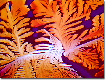
Cytidine
As one of the four building blocks (nucleosides) of nucleic acids, life as we know it would not exist without cytidine (abbreviated C by biochemists and molecular biologists). A pyrimidine nucleoside similar to thymidine (T) in DNA or uridine (U) in RNA, cytidine is an aromatic single-ring nitrogen heterocycle (cytosine) bonded to a ribose sugar unit. In nucleic acids, pyrimidine nucleosides are hydrogen bonded to purines (guanidine (G) and adenosine (A)).
View a second image of crystallized cytidine.
All of the common nucleosides are widely available in foods, but are also available to the body and its cells through biosynthetic pathways and salvage processes. Known as nitrogenized bases or monomers, nucleosides are phosphorylated and polymerized to form DNA. Cytosine, the organic base of cytidine, was first isolated from the thymus of a calf in 1894 and was synthesized in the laboratory in 1903. As a crystal, cytidine appears as a white or almost white powder and is available in a chemically synthesized regular or radiolabeled form.
When combined with phosphates and choline, cytidine 5'-diphosphocholine (CDP-choline) is prescribed for brain aging, stroke, and circulatory disorders such as ischemia, by creating an increase in membrane phospholipid turnover. Apparently, cytidine plays a role in healing and the process of tissue formation over a scab. The pyrimidine nucleoside is also indicated for therapy of a corneal inflammation or ulcer as part of an eye lotion formulation. In some recent animal experiments, antidepressant actions of cytidine were noted, with an apparent increase in mobility. Through the Human Genome Project, the gene for cytidine deaminase was mapped and delineated and the enzyme was also isolated from the common gut bacteria, Escherichia coli. The enzyme is used by geneticists and gene therapists for cutting and splicing DNA and RNA segments, and may explain somatic mutations and evolution by viral and bacterial vectors.
Contributing Authors
Omar Alvarado, Thomas J. Fellers and Michael W. Davidson - National High Magnetic Field Laboratory, 1800 East Paul Dirac Dr., The Florida State University, Tallahassee, Florida, 32310.
BACK TO THE POLARIZED LIGHT IMAGE GALLERY
BACK TO THE DIGITAL IMAGE GALLERIES
Questions or comments? Send us an email.
© 1995-2025 by Michael W. Davidson and The Florida State University. All Rights Reserved. No images, graphics, software, scripts, or applets may be reproduced or used in any manner without permission from the copyright holders. Use of this website means you agree to all of the Legal Terms and Conditions set forth by the owners.
This website is maintained by our
Graphics & Web Programming Team
in collaboration with Optical Microscopy at the
National High Magnetic Field Laboratory.
Last Modification Friday, Nov 13, 2015 at 02:19 PM
Access Count Since September 17, 2002: 12214
Visit the website of our partner in introductory microscopy education:
|
|