
Polarized Light Digital Image Gallery
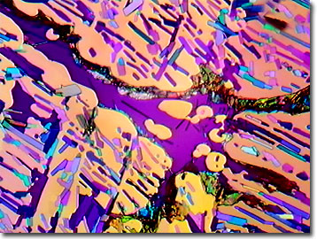
Cresyl Violet
Cresyl violet is a synthetic dye that is widely utilized to stain neuronal tissues. Because it is a basic stain, it readily binds to the acidic components of the neuronal cytoplasm such as RNA-rich ribosomes, as well as the nuclei and nucleoli of the nerve cells. Cresyl violet should not be confused with crystal violet, a stain for distinguishing Gram positive and Gram negative bacteria.
Cresyl violet is probably the third most common stain used in preparing histological sections. Marketed also as Certistain®, cresyl violet acetate appears as green crystal that melt at 142 degrees Celsius and is chemically known as aminonaptho-aminophenoxazonium acetate featuring 18 carbon, 15 hydrogen, 3 nitrogen, and 3 oxygen atoms per molecule. Another formulation, cresyl violet perchlorate, features a peak absorption wavelength of about 590 nanometers and a peak fluorescence emission wavelength of about 615 nanometers. There is some confusion in the historic histological literature over several dyes bearing the names cresyl violet, cresyl echt violet, and cresyl violet acetate based on importations of several products that are most certainly chemically related, but not exactly of the same molecular structure. There have been several descriptions of experiments using cresyl violet hydrochloride (cresyl echt violet) in the preparation of mosquito chromosome spreads and cresyl fast violet for brain and spinal tissue preparations.
When luxol fast blue, with its affinity for lipids, is coupled with cresyl violet, neuron myelin stains blue, red blood cells turquoise, and the nuclei and Nissl bodies (primarily rough endoplasmic reticulum) stain as purple. Alternatively, when nerve tissue is stained using the cresyl fast violet technique, the Nissl bodies, nuclear membranes and nucleoli stain intensely blue or violet, while the cytoplasm, ganglia cells, and glia stain weakly blue. All other cell process elements remain unstained. Some trials indicate that staining is enhanced when using fresh frozen tissues that have not been fixed. Other applications of cresyl fast violet staining include the identification of parasitic microorganisms such as Cryptococcus neoformans in their host tissues, and the staining of fixed tumor tissue. When coupled with polarized light microscopy, a spectacular birefringence is produced because of the staining of the anisotropic yeast as pink, while Blastomyces (and others) also stain pink, but are isotropic. Only Cryptococcus exhibits a pink luminous center surrounded by brilliantly birefringent spinous radiations of the thick, mucoid capsule.
Contributing Authors
Omar Alvarado, Thomas J. Fellers and Michael W. Davidson - National High Magnetic Field Laboratory, 1800 East Paul Dirac Dr., The Florida State University, Tallahassee, Florida, 32310.
BACK TO THE POLARIZED LIGHT IMAGE GALLERY
BACK TO THE DIGITAL IMAGE GALLERIES
Questions or comments? Send us an email.
© 1995-2025 by Michael W. Davidson and The Florida State University. All Rights Reserved. No images, graphics, software, scripts, or applets may be reproduced or used in any manner without permission from the copyright holders. Use of this website means you agree to all of the Legal Terms and Conditions set forth by the owners.
This website is maintained by our
Graphics & Web Programming Team
in collaboration with Optical Microscopy at the
National High Magnetic Field Laboratory.
Last Modification Friday, Nov 13, 2015 at 02:19 PM
Access Count Since September 17, 2002: 29908
Visit the website of our partner in introductory microscopy education:
|
|