Oblique Digital Image Gallery
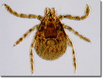
Rocky Mountain Wood Tick (Dermacentor andersoni)
The Rocky Mountain wood tick (Dermacentor andersoni) is one of two major vectors in the United States of Rocky Mountain spotted fever (Rickettsia rickettsii), a bacterial zoonosis transferred to humans from small rodents via a tick vector. Only members of the hard tick family Ixodidae are naturally infected with Rocky Mountain spotted fever, including the widespread American dog tick, D. variabilis, found east of the Rocky Mountains and in limited areas of the Pacific Coast.

Restricted in habitat to the Rocky Mountains in the United States and southwestern Canada, the Rocky Mountain wood tick's life cycle may require between two and three years for completion. Although adult ticks feed primarily on large mammals, including humans, the larval and nymph forms feast on small rodents. As with most tick species, D. andersoni requires a blood meal before developing into its next life stage. During feeding as a larva or nymph, ticks may become infected with the R. rickettsii bacteria. They pass on the infection to unsuspecting humans as adult ticks, gaining a blood meal in the bargain. Male ticks can also transfer Rocky Mountain spotted fever to female ticks through body fluids or spermatozoa during the mating ritual. Once infected, a tick can carry the pathogen for life.
Rocky Mountain spotted fever is often misdiagnosed as a case of the flu, but in its early stages, even experienced physicians have difficulty properly pin-pointing the ailment. Classically, a fever, rash, and the history of a tick bite leads to proper identification, although the characteristic red spotted rash on the palms or soles is usually not observed until after the sixth day of the symptomatic onset. Because the bacteria infects the cells lining blood vessels throughout the body, serious manifestations of Rocky Mountain spotted fever may involve the respiratory, central nervous, gastrointestinal, or renal systems. For some, within five days of onset, death follows, while others suffer permanent partial paralyses of the lower extremities, gangrene, deafness, and language disorders.
Contributing Authors
Cynthia D. Kelly, Thomas J. Fellers and Michael W. Davidson - National High Magnetic Field Laboratory, 1800 East Paul Dirac Dr., The Florida State University, Tallahassee, Florida, 32310.
BACK TO THE OBLIQUE IMAGE GALLERY
BACK TO THE DIGITAL IMAGE GALLERIES
Questions or comments? Send us an email.
© 1995-2025 by Michael W. Davidson and The Florida State University. All Rights Reserved. No images, graphics, software, scripts, or applets may be reproduced or used in any manner without permission from the copyright holders. Use of this website means you agree to all of the Legal Terms and Conditions set forth by the owners.
This website is maintained by our
Graphics & Web Programming Team
in collaboration with Optical Microscopy at the
National High Magnetic Field Laboratory.
Last Modification Friday, Nov 13, 2015 at 01:19 PM
Access Count Since September 17, 2002: 18997
Visit the website of our partner in introductory microscopy education:
|
|
