
Oblique Digital Image Gallery
Hog Louse (Haematopinus suis)
The hog louse, Haematopinus suis, is an insect that is usually four to six millimeters long and is grayish-brown with dark brown and black markings. As a chewing louse, the hog louse has a wide head necessary to support the relatively large mandibles that are an essential component of its mouthparts. The male is slightly smaller than the female hog louse and is characterized by a black streak on its ventral surface.
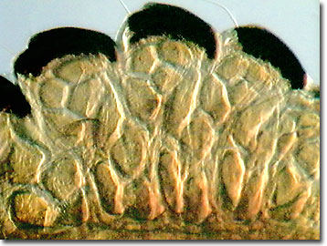
Known worldwide wherever domestic or wild swine occur, hog lice are reported to infest only swine. Although they enjoy relatively small blood meals, hog lice irritate their hosts by feeding frequently. The infected swine rub themselves to the point of injuring their skin and displacing their body hair. Severe cases result in virtually bald hogs and, with young pigs, may result in arrested growth and lost production.
Unlike most parasites, the entire life cycle of the hog louse is spent on the host. Although adult lice concentrate in the folds of the neck and jowls, at the base of the ears, inside of the legs, and on the flanks and backs of the hogs, nymphs remain concentrated on the head region. After a blood meal and mating, the female lays three to six eggs per day until she has released about 90 eggs in total. The amber eggs are deposited along the lower half of the hog's sides, on the back of the ears, the neck, shoulders, and flanks. The nits or eggs hatch in about two weeks and then the young lice molt and move to tender areas of the body to feed. Two weeks later these hog lice are mature and ready to commence the entire life cycle again. Impressively, six or more generations of hog lice may be completed with a year. Because there are neither free-living stages nor other vectors for infection, hog louse control is rather successful using insecticides and quarantines.
Contributing Authors
Cynthia D. Kelly, Thomas J. Fellers and Michael W. Davidson - National High Magnetic Field Laboratory, 1800 East Paul Dirac Dr., The Florida State University, Tallahassee, Florida, 32310.
BACK TO THE OBLIQUE IMAGE GALLERY
BACK TO THE DIGITAL IMAGE GALLERIES
Questions or comments? Send us an email.
© 1995-2025 by Michael W. Davidson and The Florida State University. All Rights Reserved. No images, graphics, software, scripts, or applets may be reproduced or used in any manner without permission from the copyright holders. Use of this website means you agree to all of the Legal Terms and Conditions set forth by the owners.
This website is maintained by our
Graphics & Web Programming Team
in collaboration with Optical Microscopy at the
National High Magnetic Field Laboratory.
Last Modification Friday, Nov 13, 2015 at 02:19 PM
Access Count Since September 17, 2002: 16708
Visit the website of our partner in introductory microscopy education:
|
|