
Oblique Digital Image Gallery
Ctenoid Fish Scales
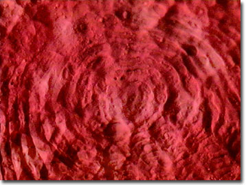
Derived from the Greek word cteno, meaning "comb", the ctenoid fish scale name refers to comb-like ctenii, which decorate the edges of the scale, as illustrated in the high magnification MIC-D digital image presented below. The spiny posterior margin of ctenoid scales has a wide variety of morphologies.
View a low magnification image of a ctenoid fish scale.
View a medium magnification image of a ctenoid fish scale.
These scales are commonly found in the majority of bony fishes (referred to as the Teleostei), and the anterior (or front) part of each scale is usually tucked behind the rear portion of the preceeding scale. As the fish grows, so do the scales, resulting in a pattern of concentric growth "rings" that increase in number with the scale size and appear similar to those found in the cross section of tree trunks. In some cases, ctenoid scale growth patterns are utilized to determine the age of a fish.
Ctenoid scales are composed of two main regions, a surface layer having a hard shell resulting from crystallized calcium salts, and a deeper layer that is built with collagen-type fibrils. Three generalized types of ctenoid scales have been categorized. They include crenate scales having simple indentations on the edges, spinoid scales that are clustered with a network of spines attached to the main scale body, and ctenoid scales that sport ctenii formed as separate bony growths, which are distinct from the scale main body.
Contributing Authors
Cynthia D. Kelly, Thomas J. Fellers and Michael W. Davidson - National High Magnetic Field Laboratory, 1800 East Paul Dirac Dr., The Florida State University, Tallahassee, Florida, 32310.
BACK TO THE OBLIQUE IMAGE GALLERY
BACK TO THE DIGITAL IMAGE GALLERIES
Questions or comments? Send us an email.
© 1995-2025 by Michael W. Davidson and The Florida State University. All Rights Reserved. No images, graphics, software, scripts, or applets may be reproduced or used in any manner without permission from the copyright holders. Use of this website means you agree to all of the Legal Terms and Conditions set forth by the owners.
This website is maintained by our
Graphics & Web Programming Team
in collaboration with Optical Microscopy at the
National High Magnetic Field Laboratory.
Last Modification Friday, Nov 13, 2015 at 02:19 PM
Access Count Since September 17, 2002: 10706
Visit the website of our partner in introductory microscopy education:
|
|