
Pond Life Digital Movie Gallery
Philodina (Rotifera) Movies
Placed in the class Bdelloidea, which is Greek for leech, species in the genus Philodina possess two ovaries and are solely parthenogenetic. Male rotifers in this class have not been observed, and members of Philodina, referred to as the world's most common metazoans, are unique to the animal kingdom.
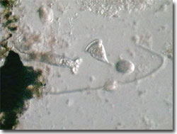
Philodina Video #1 - A rotifer extends from its attachment point to consume several items of prey; under oblique illumination with a playing time of 27 seconds. Choose a playback format that matches your connection speed:
Rotifers are found in birdbaths, ponds, puddles, roof gutters, and garden soils; the specimen illustrated here was collected from a Northern Florida pond. Up to 500 micrometers in length when extended, Philodina species are motile and do not have shells. Resembling worms, with two anterior rotating wheel organs referred to as coronas, Philodina can move like leeches or inchworms, extending and contracting as they crawl over aquatic plants and detritus. The same individuals can contract, extend their coronas, and swim freely.
Often sessile, the Philodina rotifers cement themselves to substrates while feasting on bacteria and protists. Metazoans in the family Philodinidae, these rotifers are pseudocoelomates with a body cavity containing a syncytium of amoeboid cells in a fluid. With a fixed number of cells originating in the embryo stage, Philodina rotifers grow in cell size, not cell number throughout their lives. Because of the research value of constant-cell-number organisms, Philodina are widely studied by research biologists investigating cancer processes and potential therapies.
Contributing Authors
Cynthia D. Kelly, Thomas J. Fellers and Michael W. Davidson - National High Magnetic Field Laboratory, 1800 East Paul Dirac Dr., The Florida State University, Tallahassee, Florida, 32310.
BACK TO THE DIGITAL IMAGE GALLERIES
BACK TO THE OLYMPUS MIC-D DIGITAL MICROSCOPE
Questions or comments? Send us an email.
© 1995-2025 by Michael W. Davidson and The Florida State University. All Rights Reserved. No images, graphics, software, scripts, or applets may be reproduced or used in any manner without permission from the copyright holders. Use of this website means you agree to all of the Legal Terms and Conditions set forth by the owners.
This website is maintained by our
Graphics & Web Programming Team
in collaboration with Optical Microscopy at the
National High Magnetic Field Laboratory.
Last Modification Friday, Nov 13, 2015 at 02:19 PM
Access Count Since September 17, 2002: 22383
Visit the website of our partner in introductory microscopy education:
|
|