Pond Life Digital Movie Gallery
Vorticella (Protozoa) Movies
As a member of the phylum Cilophora (ciliates), Vorticella grow in macroscopic clusters of stalked individual animals that may be mistaken for a colony of filamentous algae. Vorticella are transparent, range from 50 to 150 micrometers in length, and are easily cultured as the unicellular protist component of infusoria commonly utilized as food for fish fry.

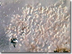
Vorticella Video #1 - A colony of ciliates moves in explosive pulses; under oblique illumination with a playing time of 32 seconds. Choose a playback format that matches your connection speed:
Often sedentary in behavior, stalked Vorticella are anchored to aquatic macrophytes, filamentous algae, and other submerged substrates. The cilia lining the "cup" of this cup-and-stalk shaped ciliate beat water currents bearing food into the cytostome for ingestion. The digestion ultimately takes place within the food vacuoles, or vesicles, that form at the end of the cytostome from detached cell membrane. Wastewater is removed by the water-expelling vesicle. When habitat conditions degrade, individuals are able to morph into motile forms, and the cilia, beating in unison, propel the unicellular animals through the water. Then en masse, the individuals migrate several inches, settle down on suitable surfaces, and grow new stalks. When disturbed or if live prey such as bacteria or protozoans is captured, the stalk can rapidly retract into a coil like a spring, through use of a full-length myoneme, a type of contractile fiber.
The u-shaped macronucleus and micronucleus of this ciliate is visible through the transparent cup. Vorticella (family Vorticellidae) reproduce, as do other related unicellular ciliates, primarily asexually by simple fission, dividing along the length of the body. However, they are also capable of sexual conjugation. In binary fission, one of the two daughter cells retains the parental stalk while the other grows temporary cilia, relocates, loses the new cilia, and subsequently grows a new stalk for attachment.
Contributing Authors
Cynthia D. Kelly, Thomas J. Fellers and Michael W. Davidson - National High Magnetic Field Laboratory, 1800 East Paul Dirac Dr., The Florida State University, Tallahassee, Florida, 32310.
BACK TO THE DIGITAL IMAGE GALLERIES
BACK TO THE OLYMPUS MIC-D DIGITAL MICROSCOPE
Questions or comments? Send us an email.
© 1995-2025 by Michael W. Davidson and The Florida State University. All Rights Reserved. No images, graphics, software, scripts, or applets may be reproduced or used in any manner without permission from the copyright holders. Use of this website means you agree to all of the Legal Terms and Conditions set forth by the owners.
This website is maintained by our
Graphics & Web Programming Team
in collaboration with Optical Microscopy at the
National High Magnetic Field Laboratory.
Last Modification Friday, Nov 13, 2015 at 01:19 PM
Access Count Since September 17, 2002: 25030
Visit the website of our partner in introductory microscopy education:
|
|
