Pond Life Digital Movie Gallery
Amoeba (Protozoa) Movies
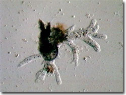
Amoebas are primitive, unicellular animals known by their flowing, free-form movements and their prey capture techniques. Most members of the order Amoebida in the phylum Sarcodina are free-living, but some are endoparasites of plants and animals, and are well known disease vectors of ailments such as amoebic dysentery in humans.

Amoeba Video #1 - Cytoplasm of an amoeba is seen flowing into the advancing pseudopods as they extend during movement; under oblique illumination with a playing time of 14 seconds. Choose a playback format that matches your connection speed:
Amoeba Video #2 - An amoeba slowly moves around the viewfield by alternately extending and retracting a pair of very long pseudopodia; under oblique illumination with a playing time of 45 seconds. Choose a playback format that matches your connection speed:
Within the order, there are the typical naked amoebas plus several shelled species. In either type, the amoeboids locomote by using their pseudopods (or false feet), which are projected in the direction of movement, and then followed by the rest of the simple organism via flowing cytoplasm (cyclosis). The best-known species is Amoeba proteus, commonly cultured and sold commercially for classroom instruction and research laboratories. In similarity to other single-celled organisms, Amoeba feature a nucleus, a contractile vacuole, a very flexible cell membrane, and cytoplasm.
When suitable prey items such as bacteria or other protists are located via chemotaxis, the pseudopods are extended and encircle the potential food. As the cytoplasm streams to the pseudopods, the phagocytosis is completed and the prey is digested and stored in the new food vacuole. Amoeboids can reproduce by binary fission and possess some powers of regeneration. In a form of asexual reproduction, the pseudopods begin by pulling apart, after which the nuclear material replicates. As the pseudopods' separation progresses, they eventually split the nucleus, and then the cell, into two smaller individuals. Recent research on combating amoebic dysentery and other amoeba-related human diseases has centered on means of stopping the reproducing amoeba from completing division. In some cases, division is not completed, and the cytoplasm rejoins to form a single individual with two nuclei. Observations in the laboratory reveal a phenomenon best described as "midwifery". As an amoeba starts to replicate, other "midwife" amoebas reacting to chemical stimuli, related to the release of sugars, lipids, and protein from the stretching cell membrane, rush to the "birthing" site. The midwife amoebas actually aid the complete division by helping in the pulling process.
Contributing Authors
Cynthia D. Kelly, Thomas J. Fellers and Michael W. Davidson - National High Magnetic Field Laboratory, 1800 East Paul Dirac Dr., The Florida State University, Tallahassee, Florida, 32310.
BACK TO THE DIGITAL IMAGE GALLERIES
BACK TO THE OLYMPUS MIC-D DIGITAL MICROSCOPE
Questions or comments? Send us an email.
© 1995-2025 by Michael W. Davidson and The Florida State University. All Rights Reserved. No images, graphics, software, scripts, or applets may be reproduced or used in any manner without permission from the copyright holders. Use of this website means you agree to all of the Legal Terms and Conditions set forth by the owners.
This website is maintained by our
Graphics & Web Programming Team
in collaboration with Optical Microscopy at the
National High Magnetic Field Laboratory.
Last Modification Friday, Nov 13, 2015 at 01:19 PM
Access Count Since September 17, 2002: 42210
Visit the website of our partner in introductory microscopy education:
|
|
