
Darkfield Digital Image Gallery
Mucor Zygotes
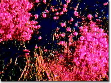
Molds of the genus Mucor are found in soil and as saprobes of plants and manure in the wild, and as a common contaminant of stored and processed foods in the kitchen. There are about 50 species described worldwide and many plague water-damaged or moist structural materials and can inflict allergies on exposed people.
View a second image of Mucor zygotes.
As a member of the phylum Zygomycota, the filamentous fungus grows rapidly at 25 to 30 degrees Celsius, sometimes several centimeters high, and at the macroscopic level, its fluffy appearance resembles cotton candy. Initially the mold is white and gradually becomes gray with maturity. The life cycle is complex and involves asexual and sexual reproductive methods. Zygospores, if present, arise from the mycelium. Species of Mucor are distinguished from one another by the branching (unbranched or branched), the presence of sporangiophores and their shape (oval or round), the maximum temperature tolerated for growth, the presence of chlamydospores, and whether or not they can assimilate ethanol. The other genera of zygomycots grow best above 40 degrees Celsius, significantly higher than the maximum of 37 degrees reported for Mucor species.
Species in the fungal genus can cause a group of infections in humans known as zygomycosis. Zygomycosis includes infections in mucous membranes, nasal passages and sinuses, eyes, lungs, skin, and brain, as well as renal and pulmonary infections and septic arthritis. People suffering from diabetes, extensive burns, immunosuppression symptoms associated with AIDS and other afflictions, or those who are intravenous drug users, appear to be most susceptible to Mucor infections. Treatments of Mucor and other fungal infections of the phylum Zygomycota are difficult because of their property of invading vascular tissues of immuno-suppressed people, and mortality rates are relatively high.
Contributing Authors
Cynthia D. Kelly, Thomas J. Fellers and Michael W. Davidson - National High Magnetic Field Laboratory, 1800 East Paul Dirac Dr., The Florida State University, Tallahassee, Florida, 32310.
BACK TO THE DARKFIELD IMAGE GALLERY
BACK TO THE DIGITAL IMAGE GALLERIES
Questions or comments? Send us an email.
© 1995-2025 by Michael W. Davidson and The Florida State University. All Rights Reserved. No images, graphics, software, scripts, or applets may be reproduced or used in any manner without permission from the copyright holders. Use of this website means you agree to all of the Legal Terms and Conditions set forth by the owners.
This website is maintained by our
Graphics & Web Programming Team
in collaboration with Optical Microscopy at the
National High Magnetic Field Laboratory.
Last Modification Friday, Nov 13, 2015 at 02:19 PM
Access Count Since September 17, 2002: 9939
Visit the website of our partner in introductory microscopy education:
|
|