
Darkfield Digital Image Gallery
Deer Tick (Ixodes dammini) Larvae
Throughout the woods of North America, the tiny deer tick (Ixodes dammini) is rapidly replacing the mythical "Boogeyman" and the fabled big, bad wolf as a means of imparting caution and obedience to children, in the face of fear. Since the 1980s, nymphal and adult deer ticks have acted as the vector for Lyme disease in North America.
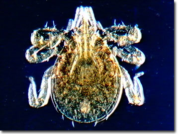
The adult deer tick is described as the size of a sesame seed (2.5-millimeter diameter), oval, and with a flattened body before enjoying a blood meal. When engorged, the eight-legged arthropod is about the size of a small pea and blue-black in color.
The etiological agent of Lyme disease, named for Lyme, Connecticut, the type locality for the zoonosis, is the spirochete Borrelia burgdorferi. This member of the hard tick family of arachnids spreads the pathological bacteria from infected host to other hosts with the larval or nymphal host, a rodent, acting as a reservoir, and an infected tick carrying the spirochetes its entire life. Some deer ticks can also cause tick paralysis and the protozoan infection caused by Babesia microti. Lyme disease is characterized by arthritis, cardiac, and skin symptoms and is also reported in dogs. Migrating birds bitten by an infected tick are believed to help spread deer ticks and their zoonoses.
In its three-step process of parasitism, larval deer ticks feed on the ears of white-footed mice while nymphs attach to humans, dogs, horses, rodents, and cattle. Adults, as the name implies, feed on the preferred white-tailed deer, as well as humans and other mammals. Blood-engorged females lay approximately 3,000 eggs and the larvae, nymphs, and adults feed on separate hosts. The entire life cycle takes about two years to complete.
Contributing Authors
Cynthia D. Kelly, Thomas J. Fellers and Michael W. Davidson - National High Magnetic Field Laboratory, 1800 East Paul Dirac Dr., The Florida State University, Tallahassee, Florida, 32310.
BACK TO THE DARKFIELD IMAGE GALLERY
BACK TO THE DIGITAL IMAGE GALLERIES
Questions or comments? Send us an email.
© 1995-2025 by Michael W. Davidson and The Florida State University. All Rights Reserved. No images, graphics, software, scripts, or applets may be reproduced or used in any manner without permission from the copyright holders. Use of this website means you agree to all of the Legal Terms and Conditions set forth by the owners.
This website is maintained by our
Graphics & Web Programming Team
in collaboration with Optical Microscopy at the
National High Magnetic Field Laboratory.
Last Modification Friday, Nov 13, 2015 at 02:19 PM
Access Count Since September 17, 2002: 15153
Visit the website of our partner in introductory microscopy education:
|
|