
Darkfield Digital Image Gallery
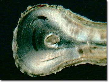
Human Tooth Root
As the anchor to a human tooth, the root extends into the bone of the jaw and houses the nerves and blood vessels that carry nutrients to the tooth. A tough, yellowish bone-like tissue, cementum, covers the root and helps hold the tooth in its socket.
View a second image of a human tooth root.
The cementum contains the periodontal membrane and is in contact with the gum tissue that separates the tooth from the underlying bone. Fibers of the periodontal membrane are embedded in the cementum. The root end openings allow passage of the neural and circulatory elements out of the tooth. Each human tooth has from one to four root canals that carry veins, arteries, lymph vessels, and nerves to the living pulp of the tooth.
For those suffering severe tooth pain and decay and facing the dreaded root canal surgery, there is no doubt that human teeth, including the roots, contain nerves. When the pulp in the root canal becomes infected due to a deep cavity or fracture, bacteria seep in and the root can die. With the damaged or dead pulp in the root, there is increased blood flow and cellular activity for attempted repairs, creating a pressure within the tooth that cannot be relieved. Severe pain in the tooth is sensed when biting down, chewing with the diseased tooth, or applying heat or cold, such as by drinking hot coffee or eating a frozen dessert. Root canal work involves severing the neural and circulatory elements, cleaning out the infected canal, and sealing it off from further infections. Without the endodontic surgery, the infection will spread, the jawbone will begin to degenerate, and the tooth will fall out. Infections in root canals of teeth are common and sometimes they become toxic, requiring tooth extraction. New research is exploring replacing decayed tooth pulp above the root as an alternative to root canal surgery.
Molecular biologists and geneticists are exploring how the tooth root is laid down and are trying to harness the natural processes to aid in reconstructive surgery and to treat genetic disorders such as cleft palate. The ultimate goal for dentists will be the ability to regenerate lost or diseased teeth instead of replacement with dentures and dental implants. As part of the Human Genome Project, the genes controlling cementum and root development have been elucidated and now researchers are exploring what mechanisms turn on and off these genes.
Comparative morphological studies of the tooth root structures in primates including humans provide clues to dietary adaptations of ancestral and extant primates. Using biomechanics, tooth roots must resist the loads applied to the tooth crown, and their various forms appear to be adapted to the varying loads experienced by different teeth within the mouth of a primate. Variation among primate species suggests different dietary histories over evolutionary time. Inferences from this work have been used to discern ancestral feeding patterns based on fossil hominid skulls. Forensic scientists are exploring the use of root coloration as a method of aging a victim with a high correlation found between increasing chronological age and increased root coloration.
Contributing Authors
Cynthia D. Kelly, Thomas J. Fellers and Michael W. Davidson - National High Magnetic Field Laboratory, 1800 East Paul Dirac Dr., The Florida State University, Tallahassee, Florida, 32310.
BACK TO THE DARKFIELD IMAGE GALLERY
BACK TO THE DIGITAL IMAGE GALLERIES
Questions or comments? Send us an email.
© 1995-2025 by Michael W. Davidson and The Florida State University. All Rights Reserved. No images, graphics, software, scripts, or applets may be reproduced or used in any manner without permission from the copyright holders. Use of this website means you agree to all of the Legal Terms and Conditions set forth by the owners.
This website is maintained by our
Graphics & Web Programming Team
in collaboration with Optical Microscopy at the
National High Magnetic Field Laboratory.
Last Modification Friday, Nov 13, 2015 at 02:19 PM
Access Count Since September 17, 2002: 17394
Visit the website of our partner in introductory microscopy education:
|
|