
Darkfield Digital Image Gallery
Chicken Embryos
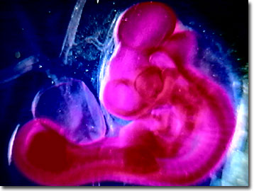
The chick or baby chicken is not only a favorite as a reminder of spring and as a live Easter gift for children in the United States, but is also well studied by embryologists, toxicologists, and histologists. Because of their relatively low cost, abundance, and year-round supply, chicken embryos make ideal laboratory organisms. The digital image presented below illustrates a darkfield microscopy view of a 80-hour chicken embryo.
View a chicken embryo that is 72 hours old.
View a chicken embryo that is 96 hours old.
Chicken embryos and cells grown in the laboratory may reduce animal testing by replacing exposure trials on mice and rats. In the field of genetic engineering, state-of-the-art research on gene function is being carried out on cultured chicken cells instead of engineered mice. Most chicken genes have equivalents in humans, therefore, utilizing chicken cells to identify the proteins for which genes code, and their locations on the chromosomes has a direct impact on understanding human biology.
Chicken embryos develop in eggs rather than inside the mother, as in mice and rats, making the control of experiments much easier for research scientists utilizing the chicken. Since 1967, when killed measles virus-derived vaccines were abandoned because of severe reactions in some children, measles vaccines are produced by weakening the live virus through transplantation from 40 times to 85 times into chicken embryos. Similarly, a stronger mumps vaccine was developed by passing the virus repeatedly through hens' eggs and chicken embryos.
Contributing Authors
Cynthia D. Kelly, Thomas J. Fellers and Michael W. Davidson - National High Magnetic Field Laboratory, 1800 East Paul Dirac Dr., The Florida State University, Tallahassee, Florida, 32310.
BACK TO THE DARKFIELD IMAGE GALLERY
BACK TO THE DIGITAL IMAGE GALLERIES
Questions or comments? Send us an email.
© 1995-2025 by Michael W. Davidson and The Florida State University. All Rights Reserved. No images, graphics, software, scripts, or applets may be reproduced or used in any manner without permission from the copyright holders. Use of this website means you agree to all of the Legal Terms and Conditions set forth by the owners.
This website is maintained by our
Graphics & Web Programming Team
in collaboration with Optical Microscopy at the
National High Magnetic Field Laboratory.
Last Modification Friday, Nov 13, 2015 at 02:19 PM
Access Count Since September 17, 2002: 11508
Visit the website of our partner in introductory microscopy education:
|
|