
Darkfield Digital Image Gallery
Aurelia Ephyra-Stage Jellyfish
Members of the jellyfish in the class Scyphozoa undergo a complex life cycle including a larval medusa or ephyra stage. The genus Aurelia demonstrates the typical life stages for this class of invertebrates, which also include the free-swimming planula, the stalked and tentacled schyphistoma polyp, the strobila bud, and the mature medusa stages.
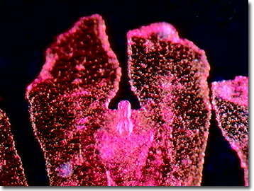
Between the dominant adult medusa stage and the small and inconspicuous larval polyp stage, Aurelia jellyfishes undergo the ephyra stage. The common and cosmopolitan moon jelly (A. aurita) is often used as a laboratory experimental and dissection model to illustrate the typical Aurelia life stages. Tiny ephyrae swim free of the sessile schyphistoma polyps. As a dispersal stage, the ephyra allows the jellyfish to spread to potential suitable habitats and prevents the placing of all offspring within one locality where they are subject to changes in environmental conditions and catastrophic events.
In less than a year, the ephyrae mature sexually into medusas. Although not mature, most of the adult characteristics (except gonads) are present in the larval ephyra stage, but have different relative sizes. Most notably, the eight pairs of lappets are disproportionately large in comparison with adult jellyfish of the same species. While lappets are inconspicuous on adult Aurelia members, they dominate the ephyrae. Other body parts such as the manubrium and the arms are common to both larval ephyrae and mature medusas in the Scyphozoa species. The rhopalia are eight sensory organs that are evenly spaced around the fringe of the bell of both stages. Each rhopalium contains a statocyst for balance and sensing gravity and an ocellus for photoreception. In the adult stage, the large lappets of the ephyra are reduced to small, triangular sense organs located adjacent to the rhopalia.
Contributing Authors
Cynthia D. Kelly, Thomas J. Fellers and Michael W. Davidson - National High Magnetic Field Laboratory, 1800 East Paul Dirac Dr., The Florida State University, Tallahassee, Florida, 32310.
BACK TO THE DARKFIELD IMAGE GALLERY
BACK TO THE DIGITAL IMAGE GALLERIES
Questions or comments? Send us an email.
© 1995-2025 by Michael W. Davidson and The Florida State University. All Rights Reserved. No images, graphics, software, scripts, or applets may be reproduced or used in any manner without permission from the copyright holders. Use of this website means you agree to all of the Legal Terms and Conditions set forth by the owners.
This website is maintained by our
Graphics & Web Programming Team
in collaboration with Optical Microscopy at the
National High Magnetic Field Laboratory.
Last Modification Friday, Nov 13, 2015 at 02:19 PM
Access Count Since September 17, 2002: 16985
Visit the website of our partner in introductory microscopy education:
|
|