
Darkfield Digital Image Gallery
Mold (Aspergillus) Conidiophores
As one of the two common genera of molds on fruits and other foods such as grain, wheat, and bread, species of the genus Aspergillus are distinguished from Penicillium species by the origin of their spore-bearing stalks or conidiophores. In Aspergillus, the conidiophore arises from a foot-cell, a vegetative mycelium.
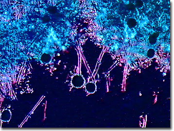
The foot-cell is broader than the filament of the mycelium, and when the spores are ripe, it usually does not contain protoplasm. At the end of the non-septate (no cross walls) stalk, a swollen vesicle bears a large number of little stalks or sterigmata. Secondary branches or sterigmata bear the numerous spores.
Aspergillus species are common, widespread, and occur naturally in soils and on plant matter. Most of the species, including the most ubiquitous A. niger, prefer relatively warm habitats ranging from 35 to 40 degrees Celsius for an optimal temperature. In microbiological and tissue culture laboratories, this species is one of the most troublesome as a contaminating mold. Several human diseases are tied to inhaling Aspergillus spores, especially if the person is suffering from an impaired immune system and the possibility of forming mycotoxins.
Not all Aspergillus molds are considered a nuisance to humans. In fact, the koji mold (A. oryzae) produces kojic acid, a metabolite that possesses antibacterial and antifungal properties and is very important in Japan for the production of sake (rice wine), miso (soybean paste), shoyu (soy sauce), amasake (sweet rice milk), and shouchu (distilled rice, brown sugar, or sweet potato liquor, which is similar to brandy).
Contributing Authors
Cynthia D. Kelly, Thomas J. Fellers and Michael W. Davidson - National High Magnetic Field Laboratory, 1800 East Paul Dirac Dr., The Florida State University, Tallahassee, Florida, 32310.
BACK TO THE DARKFIELD IMAGE GALLERY
BACK TO THE DIGITAL IMAGE GALLERIES
Questions or comments? Send us an email.
© 1995-2025 by Michael W. Davidson and The Florida State University. All Rights Reserved. No images, graphics, software, scripts, or applets may be reproduced or used in any manner without permission from the copyright holders. Use of this website means you agree to all of the Legal Terms and Conditions set forth by the owners.
This website is maintained by our
Graphics & Web Programming Team
in collaboration with Optical Microscopy at the
National High Magnetic Field Laboratory.
Last Modification Friday, Nov 13, 2015 at 02:19 PM
Access Count Since September 17, 2002: 40977
Visit the website of our partner in introductory microscopy education:
|
|