Darkfield Digital Image Gallery
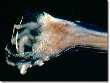
Canine Hookworm
Hookworm infestation, known also as ancylostomiasis, is the most common and most serious endoparasitic infection of dogs in the tropics, and affects cats as well, although to a lesser extent. Adult hookworms attach to small arteries within the intestines of the host and take blood meals directly. As a truly prolific egg producer, the female canine hookworm lays up to 30,000 ova per day, which are then passed into the environment via the animal's droppings.

View a second image of a canine hookworm.
The canine hookworm larvae hatch in soil where they develop and await a passing host that is susceptible to infestation. A dog may ingest soil containing the larvae, or more commonly, the larval hookworms enter the animal by penetrating the paws. Over time, the larvae migrate within the body to the lungs, ascend the respiratory tract, and eventually are swallowed. They move through the digestive system until they reach the lining of the small intestine, where they attach and feed, starting the cycle once again.
With a typical three-week life cycle, infestation becomes particularly problematic in tropical or sub-tropical environs. Dogs suffering from hookworm loadings in their guts often experience weight loss, diarrhea, black and tarry stools, and severe anemia. Significant infestations may be fatal for puppies. Unlike some other hookworm species and other endoparasites, the canine hookworm can also infest humans.
Contributing Authors
Cynthia D. Kelly, Thomas J. Fellers and Michael W. Davidson - National High Magnetic Field Laboratory, 1800 East Paul Dirac Dr., The Florida State University, Tallahassee, Florida, 32310.
BACK TO THE DARKFIELD IMAGE GALLERY
BACK TO THE DIGITAL IMAGE GALLERIES
Questions or comments? Send us an email.
© 1995-2025 by Michael W. Davidson and The Florida State University. All Rights Reserved. No images, graphics, software, scripts, or applets may be reproduced or used in any manner without permission from the copyright holders. Use of this website means you agree to all of the Legal Terms and Conditions set forth by the owners.
This website is maintained by our
Graphics & Web Programming Team
in collaboration with Optical Microscopy at the
National High Magnetic Field Laboratory.
Last Modification Friday, Nov 13, 2015 at 01:19 PM
Access Count Since September 17, 2002: 16411
Visit the website of our partner in introductory microscopy education:
|
|
