
Brightfield Digital Image Gallery
Starfish Brachiolaria Larva
Larval specimens of the common starfish Asterias rubens (phylum Echinodermata) occur in the plankton among those of sea urchins, sea cucumbers, and brittle stars. A majority of the starfish species disperse exceedingly large numbers of their gametes (eggs and sperm) freely into the water in the hope of an external fertilization event.
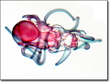
In order to increase the chance of fertilization, it is believed that starfish gather in groups when they are ready to spawn, employing several environmental signals to coordinate the timing. These include the length of the day to indicate the appropriate time of year, and the occurance of dawn or dusk to determine the correct time of day. Starfish may also use chemical signals to initiate the fertilization process. After the gametes are matched and undergo fertilization, a blastula develops that employs cilia to enable free swimming of the embryo. The next stage is the gastrula, followed by differentiation into the bipinnaria larval stage. Several more weeks into the developmental period, the brachiolaria larval stage is reached. These larvae have a gut containing internal cilia, which the organism utilizes to inhale and transport food particles, such as diatoms and other microscopic creatures in the plankton.
Brachiolaria larvae live as plankton, suspended in the ocean water and moving about by a coordinated motion of the external cilia. The larvae are bilaterally symmetrical and have a distinct left and right side (unlike adults of the species, which have a radial symmetry). Eventually, the larvae undergo a complete metamorphosis, settle to the bottom of the sea, and slowly grow into adults. The entire process, from egg to mature starfish, takes about two months depending upon the prevailing water temperature. After about a year, the newly formed starfish is ready to participate in the reproductive chain.
Contributing Authors
Cynthia D. Kelly, Thomas J. Fellers and Michael W. Davidson - National High Magnetic Field Laboratory, 1800 East Paul Dirac Dr., The Florida State University, Tallahassee, Florida, 32310.
BACK TO THE BRIGHTFIELD IMAGE GALLERY
BACK TO THE DIGITAL IMAGE GALLERIES
Questions or comments? Send us an email.
© 1995-2025 by Michael W. Davidson and The Florida State University. All Rights Reserved. No images, graphics, software, scripts, or applets may be reproduced or used in any manner without permission from the copyright holders. Use of this website means you agree to all of the Legal Terms and Conditions set forth by the owners.
This website is maintained by our
Graphics & Web Programming Team
in collaboration with Optical Microscopy at the
National High Magnetic Field Laboratory.
Last Modification Friday, Nov 13, 2015 at 02:19 PM
Access Count Since September 17, 2002: 23610
Visit the website of our partner in introductory microscopy education:
|
|