
Brightfield Digital Image Gallery
Primate Ileum Stained Thin Section
The ileum, the lowest section of the small intestine, connects to the jejunum and the large intestine. In common with other portions of the primate small intestine, the ileum employs small finger-like appendages, or villi, for the absorption of nutrients from digesting matter.
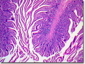
Each villus is composed of large, circular folds of the small intestine, a capillary bed, a lacteal (lymphatic capillary), smooth muscle, and a brush border (microvilli). The microvilli complete the digestion of carbohydrates into monosaccharides, and the remaining small peptides into amino acids. The products of this digestion are absorbed efficiently by the microvilli into the circulating blood because their geometry maximizes the surface area for enzyme secretion and nutrient absorption. The lacteal system (lymph vessels) of the ileum absorbs, as globules, the glycerol, fatty acids, and triglycerides of digested fats and oils into the large veins of the circulatory system that drain into the heart. Interestingly, this means that the products of fat digestion enter directly into the bloodstream while digested protein, carbohydrates, nucleic acids, vitamins, and minerals are actively absorbed by the ileum's capillary blood system within the villi. These non-fat digestion products are then transported via the hepatic portal vessel to the liver for further processing. The villi, with the help of the mucosa (the lining of the lumen), also aid the movement of the bolus, complete with solid waste in the form of fecal matter, into the large intestine for compaction and elimination by the rectum.
The composition of the mucosa is primarily simple columnar epithelium, but it is rich in mucus-secreting goblet cells, and includes a scant, smooth muscle layer (muscularis mucosae). It aids digestion by secreting digestive enzymes and hormones. The lamina propria consists of loose areolar connective tissue in the ileum's mucosa that contains isolated lymph nodes for disease fighting, in addition to capillaries that nourish epithelium and absorb digested nutrients. External to the mucosa, the submucosa of the ileum is made up of connective tissue that contains the blood and lymph vessels, lymph nodes, nerve fibers, and elastic fibers. As part of the autonomic nervous system, the submucosal plexus is an intrinsic nerve supply of the gastrointestinal tract and helps control the activity of glands and smooth muscle. The next layer of the ileum, the muscularis externa, is composed of an inner circular layer and outer longitudinal layer of smooth muscle that, through peristalsis, propels and mixes the digesting food matter. Paneth cells within the small intestine's ileum secrete lysozyme.
The gastrointestinal tract of nonhuman primates is dominated by the colon, while humans have a more prominent small intestine (21 feet long), both in volume and in length, when compared proportionally to the rest of the body. This divergence between humans and some of the great apes, such as gorillas and orangutans, reflects vast differences in diet, with omnivorous humans depending on higher quality (more protein, fats, carbohydrates) food. The slower moving, nonhuman vegetarian primates eat lower quality plants with a higher proportion of roughage. In humans, the lining of the small intestine has a surface area of about 300 square meters, approximately the area of a regulation tennis court, and the average transit time for digesting food is three to six hours. The ileum of the typical human adult is 12 feet long compared to the 10-inch duodenum and 8-foot long jejunum.
Contributing Authors
Cynthia D. Kelly, Thomas J. Fellers and Michael W. Davidson - National High Magnetic Field Laboratory, 1800 East Paul Dirac Dr., The Florida State University, Tallahassee, Florida, 32310.
BACK TO THE BRIGHTFIELD IMAGE GALLERY
BACK TO THE DIGITAL IMAGE GALLERIES
Questions or comments? Send us an email.
© 1995-2025 by Michael W. Davidson and The Florida State University. All Rights Reserved. No images, graphics, software, scripts, or applets may be reproduced or used in any manner without permission from the copyright holders. Use of this website means you agree to all of the Legal Terms and Conditions set forth by the owners.
This website is maintained by our
Graphics & Web Programming Team
in collaboration with Optical Microscopy at the
National High Magnetic Field Laboratory.
Last Modification Friday, Nov 13, 2015 at 02:19 PM
Access Count Since September 17, 2002: 17787
Visit the website of our partner in introductory microscopy education:
|
|