Brightfield Digital Image Gallery
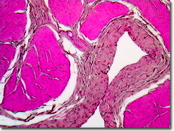
Primate Bladder
In the urinary tract system that filters the blood to produce waste urine, the primate urinary bladder is a principal component. The bladder is a muscular, balloon-shaped organ that stores urine, and at the appropriate time, squeezes it out via the urethra.

The ureters, tubes that carry the urine, connect the bladder to the kidneys, which sit at the top of the urinary tract. Nested in the pelvis, the bladder connects to the ureters via the ureterovesical junction, and their walls act as flap valves, preventing backflow of urine. Unlike the thin-walled ureters and renal pelvis, the bladder is a thick-walled structure with a heavy muscular coat that is surrounded by a layer of fat. The bladder expands and contracts based on the volume of fluid contained. In primates, the control of bladder contraction lies in the domain of the brain and spinal cord neurons, and involves both voluntary and reflex reactions. The lamina propria is a specialized layer of blood vessels and cells, which separates the lining layer from the actual bladder musculature.
Nonhuman primates have been used as research subjects for pharmacological trials to assess the efficacy of prescribed treatments for human bladder cancer and to explore the physiology of the healthy primate bladder. Applying the latest in tissue culture and cloning techniques, artificial bladders grown in primate laboratories have successfully sustained dogs for more than a year, lending promise to future human bladder repairs and transplants using simian-derived tissues and organs.
Contributing Authors
Cynthia D. Kelly, Thomas J. Fellers and Michael W. Davidson - National High Magnetic Field Laboratory, 1800 East Paul Dirac Dr., The Florida State University, Tallahassee, Florida, 32310.
BACK TO THE BRIGHTFIELD IMAGE GALLERY
BACK TO THE DIGITAL IMAGE GALLERIES
Questions or comments? Send us an email.
© 1995-2025 by Michael W. Davidson and The Florida State University. All Rights Reserved. No images, graphics, software, scripts, or applets may be reproduced or used in any manner without permission from the copyright holders. Use of this website means you agree to all of the Legal Terms and Conditions set forth by the owners.
This website is maintained by our
Graphics & Web Programming Team
in collaboration with Optical Microscopy at the
National High Magnetic Field Laboratory.
Last Modification Friday, Nov 13, 2015 at 01:19 PM
Access Count Since September 17, 2002: 11991
Visit the website of our partner in introductory microscopy education:
|
|
