
Brightfield Digital Image Gallery
Staminate Pine Cone
Male (staminate) pine cones are very small in size, ranging from 1 to 2 centimeters in diameter, and are produced on the same tree as the ovulate cones. The image presented below, captured at high magnification with the MIC-D digital microscope, is a stained thin section of staminate cone tissue, revealing an array of winged pollen grains within the fertile leaves.
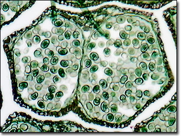
The anatomy of a staminate pine cone is structured around a spiral series of microsporophylls (the fertile male leaves) that each bear two microsporangia on the lower surface. Pollen mother cells are developed within the young microsporangium. Each cell divides by the process of meiosis to form four microspores, which are arranged in a spherical cloverleaf-like structure and ultimately will produce microgametophytes (commonly referred to as pollen grains).
At the end of the development period, the microsporangia open and release their store of pollen grains into the atmosphere to begin the fertilization process. At the same time, the scales of young ovulate pine cones separate to allow the dispersed pollen grains to intercalate the scales and adhere to a viscous fluid secreted by the ovule through a small pore called the micropyle. The pollen grain germinates, forming a pollen tube that is drawn through the micropyle. The megasporangia is eventually digested by the slow-growing pollen tube. During the approximately one year growth period of the pollen tube, the megagametophyte develops through multiple mitotic divisions inside the ovule until it reaches the mature archegonium stage, when it is ready for fertilization.
Contributing Authors
Cynthia D. Kelly, Thomas J. Fellers and Michael W. Davidson - National High Magnetic Field Laboratory, 1800 East Paul Dirac Dr., The Florida State University, Tallahassee, Florida, 32310.
Sunshyne Hendrix, Department of Biology Baylor University
BACK TO THE BRIGHTFIELD IMAGE GALLERY
BACK TO THE DIGITAL IMAGE GALLERIES
Questions or comments? Send us an email.
© 1995-2025 by Michael W. Davidson and The Florida State University. All Rights Reserved. No images, graphics, software, scripts, or applets may be reproduced or used in any manner without permission from the copyright holders. Use of this website means you agree to all of the Legal Terms and Conditions set forth by the owners.
This website is maintained by our
Graphics & Web Programming Team
in collaboration with Optical Microscopy at the
National High Magnetic Field Laboratory.
Last Modification Monday, Feb 19, 2018 at 12:04 PM
Access Count Since September 17, 2002: 36077
Visit the website of our partner in introductory microscopy education:
|
|