Brightfield Digital Image Gallery
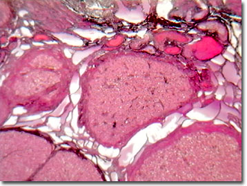
Corpus Luteum Thin Section
In the human ovary, follicles, the precursor cells of ova, undergo a complex monthly maturation cycle. At birth of a female approximately 400,000 ovarian follicles have been developed, but only about 400 enter into ovulation during a woman's childbearing years. The corpus luteum is the enlarged remnant of the secondary (Graafian) follicle following monthly ovulation, and it features vacuoles associated with the fat-soluble steroids, estrogen and progesterone.

The female sex hormone, progesterone, plays a central role in the female reproductive cycle and is produced in the adrenal glands, the placenta, and the corpus luteum of the ovary. In the absence of oocyte fertilization, progesterone levels drop and menstruation occurs. However if the egg is fertilized, progesterone levels increase and the corpus luteum is maintained, instead of shedding.
The initial follicular development occurs during the proliferative phase of the menstrual cycle, approximately 1 to 14 days from the first sign of menstrual flow, and ends at ovulation. During this phase, a small subset of the primordial follicles is stimulated by hormones to develop, and enlarges via the accumulation of follicular fluid. As the follicles develop, the granulosa layer secretes estrogen. The release of gonadotropin-releasing hormone from the pituitary gland stimulates the secretion of follicle-stimulating hormone, which is necessary in follicular development. In the human ovary, only one of the eight developing follicles develops into the dominant mature Graafian follicle and ovulates on about day 14. A feedback mechanism places the dominant follicle (greater than 11 millimeters in diameter) in the role of secreting large amounts of estrogen, which sends a message to the pituitary gland to reduce hormone stimulation, retarding the follicular development of the subordinate follicles. After expelling the oocyte, the follicle heals and its walls thicken as the cells enlarge and fill with lipids (lutenizing). The follicle then fills with blood, and becomes the corpus luteum, contributing to hormone secretion in the secretory phase. If pregnancy occurs, the trophoblast secretes human chorionic gonadotropin and the corpus luteum is maintained throughout gestation.
Functional ovarian cysts, often benign, result from a collection of fluid forming around a developing egg (follicular cyst) or in the case of the corpus luteum, a corpus luteum cyst. Although benign, these cysts are often a cause of anxiety for the women bearing them, considering the possibility of life-threatening ovarian tumors and cancer. Usually, the corpus luteum cysts resorb back into the body's fluids within a few weeks without any treatment.
Contributing Authors
Cynthia D. Kelly, Thomas J. Fellers and Michael W. Davidson - National High Magnetic Field Laboratory, 1800 East Paul Dirac Dr., The Florida State University, Tallahassee, Florida, 32310.
BACK TO THE BRIGHTFIELD IMAGE GALLERY
BACK TO THE DIGITAL IMAGE GALLERIES
Questions or comments? Send us an email.
© 1995-2025 by Michael W. Davidson and The Florida State University. All Rights Reserved. No images, graphics, software, scripts, or applets may be reproduced or used in any manner without permission from the copyright holders. Use of this website means you agree to all of the Legal Terms and Conditions set forth by the owners.
This website is maintained by our
Graphics & Web Programming Team
in collaboration with Optical Microscopy at the
National High Magnetic Field Laboratory.
Last Modification Friday, Nov 13, 2015 at 01:19 PM
Access Count Since September 17, 2002: 17417
Visit the website of our partner in introductory microscopy education:
|
|
