
Brightfield Digital Image Gallery
Obelia Hydroid
Obelia belongs to the phylum Cnidaria, which includes corals, sea anemones, jellyfish, and the freshwater hydra. The many species of this genus are widely distributed throughout all the oceans and are typical of cnidarians, both in their morphology and in their life cycle.
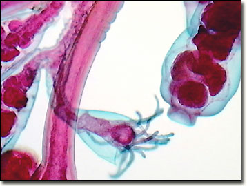
These animals take two generations to complete one life cycle. One generation, illustrated in the digital image above, and captured by the MIC-D, lives in hydroid colonies, consisting of polyps. The polyps are stalklike forms that attach to a surface (usually ocean bottom) by means of rootlike filaments. The polyps reproduce asexually, by budding, and create new polyps until a treelike colony has been formed. The colonies are dimorphic, having two polyp types. Gastrozooids, or hydranths, are the feeding polyps. They possess a mouth surrounded by stinging tentacles, giving them a flowerlike appearance, and are responsible for capturing and consuming food. Food is digested in the gastrovascular cavity and provided to the rest of the colony. Gonozooids are the reproductive polyps and, through budding, produce the next generation -- tiny jellyfish called medusae.
The second generation of the life cycle begins when the medusae are released by the gonozooids and become free-swimming forms. The medusae reproduce sexually, producing eggs and sperm that fertilize to become ciliated larvae (planulae). The planulae remain in a free-swimming form for a period of time, eventually attaching to a surface and developing into polyps.
Contributing Authors
Cynthia D. Kelly, Thomas J. Fellers and Michael W. Davidson - National High Magnetic Field Laboratory, 1800 East Paul Dirac Dr., The Florida State University, Tallahassee, Florida, 32310.
BACK TO THE BRIGHTFIELD IMAGE GALLERY
BACK TO THE DIGITAL IMAGE GALLERIES
Questions or comments? Send us an email.
© 1995-2025 by Michael W. Davidson and The Florida State University. All Rights Reserved. No images, graphics, software, scripts, or applets may be reproduced or used in any manner without permission from the copyright holders. Use of this website means you agree to all of the Legal Terms and Conditions set forth by the owners.
This website is maintained by our
Graphics & Web Programming Team
in collaboration with Optical Microscopy at the
National High Magnetic Field Laboratory.
Last Modification Friday, Nov 13, 2015 at 02:19 PM
Access Count Since September 17, 2002: 15743
Visit the website of our partner in introductory microscopy education:
|
|