
Brightfield Digital Image Gallery
Mucous Colon Epithelial Tissue
In the human gastrointestinal tract, most of the digestive system, from the mouth to the anus, follows the general scheme of hollow organs composed of four layers: the inner layer or mucosa epithelium, the submucosa, the muscularis, and the outer protective layer known as the adventitia or serosa. Following the small intestine, the large intestine includes the ascending, transverse, descending, and sigmoid sections of the colon.
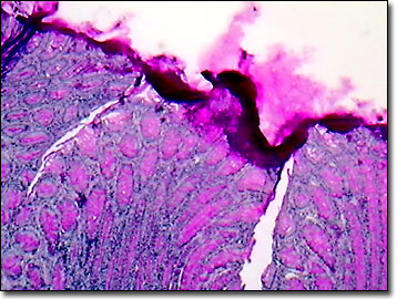
Typically, the simple epithelial tissue of the colon is made of up several different types of cells, including the characteristic goblet cells and numerous columnar absorptive cells. Named for their shape that incorporates a narrow cytoplasm base, and mucous resembling the bell of a wine goblet, goblet cells are rich with large Golgi apparatus. Unlike the preceding ileum of the small intestine, the colon does not feature villi, but has gastric pits, as does the stomach. The colon functions as the site of absorption of water, residual amino acids, lipids, carbohydrates, electrolytes, and some vitamins, while it also compacts the feces for elimination. The copious mucus produced by the goblet cells is required to facilitate passage of the dehydrated, undigested matter.
Hormones, such as vasoactive intestinal peptide, increase gut motility and stimulate intestinal ion, water, and mucus secretions, keeping food and undigested waste moving along the gastrointestinal tract. Recently, the genes that control the production and release of vasoactive intestinal protein hormone were located and mapped as part of the Human Genome Project. The bioactive peptide is now being employed in hormone therapies following cardiac surgery, menopause, and in some cancer treatments. Along with the goblet cells and columnar absorptive cells, a few enterocytes and migrating lymphocytes are found in the human colon mucosa epithelium. The crypts of Lieberkuhn, tubular glands of the colon, extend from the surface through the thickness of the mucosa.
Contributing Authors
Cynthia D. Kelly, Thomas J. Fellers and Michael W. Davidson - National High Magnetic Field Laboratory, 1800 East Paul Dirac Dr., The Florida State University, Tallahassee, Florida, 32310.
BACK TO THE BRIGHTFIELD IMAGE GALLERY
BACK TO THE DIGITAL IMAGE GALLERIES
Questions or comments? Send us an email.
© 1995-2025 by Michael W. Davidson and The Florida State University. All Rights Reserved. No images, graphics, software, scripts, or applets may be reproduced or used in any manner without permission from the copyright holders. Use of this website means you agree to all of the Legal Terms and Conditions set forth by the owners.
This website is maintained by our
Graphics & Web Programming Team
in collaboration with Optical Microscopy at the
National High Magnetic Field Laboratory.
Last Modification Friday, Nov 13, 2015 at 02:19 PM
Access Count Since September 17, 2002: 30749
Visit the website of our partner in introductory microscopy education:
|
|