
Brightfield Digital Image Gallery
Mammalian Sympathetic Ganglion
Mammalian sympathetic ganglion cells, typically obtained from laboratory rats and guinea pigs, are often used as models by physiologists to explain the biochemistry of nerve excitation and the effects that chemical substances, such as nicotine, may have on these processes. Such cells are also employed in medical research studies exploring possible solutions to devastating neuromuscular diseases such as cerebral palsy and muscular dystrophy, and potential therapies, where human experimentation is considered unethical and often illegal.
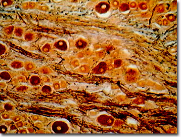
The sympathetic nervous system, the fight or flight system, allows a mammal to react rapidly by increasing blood pressure and heart beat rate, and by retarding the digestive process. Originating in the spinal cord, sympathetic nerves project to a chain of ganglia (nerve concentrations) located near the spinal cord. Usually, a synapse is made in the ganglion with another neuron, the post-ganglionic neuron, and it targets either a muscle or a gland. Mammalian sympathetic ganglia use acetylcholine as a neurotransmitter while the synapses of the target organs, and the post-ganglionic neurons, use norepinephrine.
As part of the autonomic nervous system, the sympathetic nervous system works in concert with the parasympathetic nervous system, the portion responsible for rest and digestive responses. In 1936, Sir Henry Hallett Dale received the Nobel Prize in physiology and medicine for his pioneering work describing the chemical transmission of nerve impulses, as studied with acetylcholine in the mammalian sympathetic ganglia. Playing a critical role in biological optics, ganglion cells are the final output neurons of the vertebrate retina, in which the axons of the ganglia make up the optic nerve.
Contributing Authors
Cynthia D. Kelly, Thomas J. Fellers and Michael W. Davidson - National High Magnetic Field Laboratory, 1800 East Paul Dirac Dr., The Florida State University, Tallahassee, Florida, 32310.
BACK TO THE BRIGHTFIELD IMAGE GALLERY
BACK TO THE DIGITAL IMAGE GALLERIES
Questions or comments? Send us an email.
© 1995-2025 by Michael W. Davidson and The Florida State University. All Rights Reserved. No images, graphics, software, scripts, or applets may be reproduced or used in any manner without permission from the copyright holders. Use of this website means you agree to all of the Legal Terms and Conditions set forth by the owners.
This website is maintained by our
Graphics & Web Programming Team
in collaboration with Optical Microscopy at the
National High Magnetic Field Laboratory.
Last Modification Friday, Nov 13, 2015 at 02:19 PM
Access Count Since September 17, 2002: 13438
Visit the website of our partner in introductory microscopy education:
|
|