Brightfield Digital Image Gallery
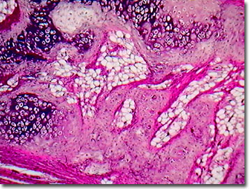
Mammalian Elastic Cartilage
Connective tissue includes bone, cartilage, tendons, and fatty tissues. Differing from articular (or hyaline) cartilage that depends on a stiff, fibrillar collagen network with fluid-filled spaces, mammalian elastic cartilage contains little or no collagen. Rather, elastic cartilage relies on other fibrillar matrix proteins for structural integrity. Elastin, the most well-studied of these proteins, forms the matrix of elastic cartilages typical of the mammalian ear, laryngeal tissues, and the epiglottis.

As one of the standard histology preparations for microscopy, mammalian elastic cartilage is often taken from laboratory rats or rabbits. Using polarizing light microscopy, the microscopist can quantitatively investigate the optical properties of isolated elastin fibers. The elastin fibers are a characteristic yellow in color and extend through the cartilage matrix in all directions. Mammalian elastic cartilage maintains the shape and flexibility of structures or organs such as the rabbit ear, while supporting and strengthening them.
As cartilage grows, mitosis takes place, but because of the matrix, the daughter cells cannot migrate. Thus, cartilage cells, or chondrocytes, tend to occur in small clusters, with each cluster representing a group of cells produced by many cellular divisions in the immediate region. Chondrocytes bear a close resemblance to fibroblasts, and in the formation of new cartilage, fibroblasts can differentiate into chondroblasts, form new cartilage, and then transform into chondrocytes, which are the maintenance cells of established cartilage tissue.
Contributing Authors
Cynthia D. Kelly, Thomas J. Fellers and Michael W. Davidson - National High Magnetic Field Laboratory, 1800 East Paul Dirac Dr., The Florida State University, Tallahassee, Florida, 32310.
BACK TO THE BRIGHTFIELD IMAGE GALLERY
BACK TO THE DIGITAL IMAGE GALLERIES
Questions or comments? Send us an email.
© 1995-2025 by Michael W. Davidson and The Florida State University. All Rights Reserved. No images, graphics, software, scripts, or applets may be reproduced or used in any manner without permission from the copyright holders. Use of this website means you agree to all of the Legal Terms and Conditions set forth by the owners.
This website is maintained by our
Graphics & Web Programming Team
in collaboration with Optical Microscopy at the
National High Magnetic Field Laboratory.
Last Modification Friday, Nov 13, 2015 at 01:19 PM
Access Count Since September 17, 2002: 19668
Visit the website of our partner in introductory microscopy education:
|
|
