Brightfield Digital Image Gallery
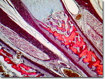
Human Fetus Skeletal Joint
Humans and other mammals incorporate joints in their skeletal structure, which allow both complex movement and a degree of flexibility that helps avoid bone breakage. Joints, which may or may not be cartilaginous, develop from mesoderm of the embryo, or in the case of the human fetus, after the ossification of the bones of the fetus. The non-condensed, undifferentiated portions of mesoderm between the bones may develop by one of three mechanisms.

View a second image of the human fetus joint.
In the case of the skull bones, the mesoderm is converted into fibrous tissue known as a synarthrodial joint. These joints may become partly cartilaginous, and are then known as amphiarthrodial joints. A third type of joint that develops in the fetus, the diathrodial joint, is recognized by its open texture and a central cavity lined with a synovial membrane.
The capsule of the joint that forms over the synovial membrane originates from tissue surrounding the original mesodermal core, which is also the source of the fibrous sheaths surrounding developing bones. In some of the movable joints, the original mesoderm between the bone ends is not completely absorbed; instead, a portion persists as the articular disk. Rupture or other damage to these capsules and disks allows friction and wearing of adjacent articulating bones, and causes pain due to the inflammation of the joint. This condition is considered a form of osteoarthritis or post-traumatic arthritis.
Contributing Authors
Cynthia D. Kelly, Thomas J. Fellers and Michael W. Davidson - National High Magnetic Field Laboratory, 1800 East Paul Dirac Dr., The Florida State University, Tallahassee, Florida, 32310.
BACK TO THE BRIGHTFIELD IMAGE GALLERY
BACK TO THE DIGITAL IMAGE GALLERIES
Questions or comments? Send us an email.
© 1995-2025 by Michael W. Davidson and The Florida State University. All Rights Reserved. No images, graphics, software, scripts, or applets may be reproduced or used in any manner without permission from the copyright holders. Use of this website means you agree to all of the Legal Terms and Conditions set forth by the owners.
This website is maintained by our
Graphics & Web Programming Team
in collaboration with Optical Microscopy at the
National High Magnetic Field Laboratory.
Last Modification Friday, Nov 13, 2015 at 01:19 PM
Access Count Since September 17, 2002: 8458
Visit the website of our partner in introductory microscopy education:
|
|
