
Brightfield Digital Image Gallery
Human Fingernail Development
Nails, whether fingernails or toenails, are derived from the same source of ectodermal skin cells in humans. Atypical, humans appear to conserve into adulthood more of the common traits that are generally shared by young primates such as predominantly naked skin (hairless) and nails that may actually be fetalized claws.
View a high magnification image of human fingernail development.
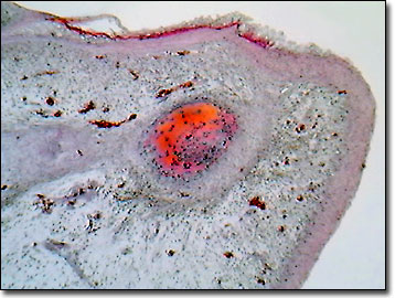
During week 10 of human development, the fetus develops its fingernails first, which are followed by the ten toenails (week 14), just as the arms and hands develop before the legs and feet. Originally, the nail fields appear at the tips of the digits and then migrate toward the dorsal surfaces. While the surrounding cells form the nail folds, keratinization of the proximal nail folds forms the nail plates. By week 32, the fingernails, and by week 36, toenails, reach the tips of the digits. Often the stage of nail growth of a baby is utilized as an indicator of the degree of prematurity.
Nails are flattened, elastic structures of a horny texture that protect the tips of the terminal phalanges (toes and fingers). Convex on the outer surface and concave within, human nails are implanted by their root into a groove in the skin (nail sulcus). The nail matrix, underlying the body and root of the nail, is the source of new nail production. The white part of the nail, the lunula, represents the portion of the nail that is not firmly attached to the connective tissue base and contrasts with the redder, highly vascularized majority of the nail that is attached to the thick matrix. Cuticles are continuous with the horny substance of the nail as part of the epidermis. Nails grow in length by producing new cells at the root of the nail, and at the distal free edge, the oldest nail cells reside.
Ectodermally derived organ abnormalities or dysplasias, such as nail defects, sparse hair or sweat glands, or congenitally missing teeth, imply developmental commonalities for these diverse parts of the human anatomy, which are derived from skin tissues. Interestingly, recent advances in molecular genetics reveal that the development of nails, kneecaps, and kidney and eye tissues, are controlled by a single gene. This realization helps explain the rare human genetic disorder that causes abnormal fingernails and prevents the development of kneecaps (nail-patella syndrome) as a single mutation.
Contributing Authors
Cynthia D. Kelly, Thomas J. Fellers and Michael W. Davidson - National High Magnetic Field Laboratory, 1800 East Paul Dirac Dr., The Florida State University, Tallahassee, Florida, 32310.
BACK TO THE BRIGHTFIELD IMAGE GALLERY
BACK TO THE DIGITAL IMAGE GALLERIES
Questions or comments? Send us an email.
© 1995-2025 by Michael W. Davidson and The Florida State University. All Rights Reserved. No images, graphics, software, scripts, or applets may be reproduced or used in any manner without permission from the copyright holders. Use of this website means you agree to all of the Legal Terms and Conditions set forth by the owners.
This website is maintained by our
Graphics & Web Programming Team
in collaboration with Optical Microscopy at the
National High Magnetic Field Laboratory.
Last Modification Friday, Nov 13, 2015 at 02:19 PM
Access Count Since September 17, 2002: 22107
Visit the website of our partner in introductory microscopy education:
|
|