
Brightfield Digital Image Gallery
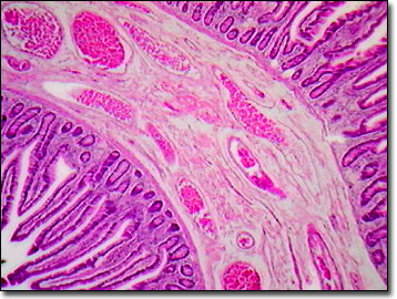
Human Lower Duodenum
The duodenum joins the jejunum and ileum as one of the three major sections of the human small intestine in the lower gastrointestinal (GI) system. To aid peristalsis (the movement of digesting matter), there are inner circular and outer longitudinal layers of smooth muscle surrounding the duodenum.
View a second image of human lower duodenum.
Characteristic of the duodenum or upper small intestine, Brunner's glands are situated in the submucosa as are Meissner's nerve plexuses. The highly alkaline mucus secretions from the Brunner's glands serve to change the acidic chyme of the stomach to a basic pH, furthering the digestive processes. These glandular secretions pass through the muscularis mucosa via ducts, on their way to the lumen of the duodenum.
Outside of the muscularis mucosa layer, the muscularis externa layer is composed of two smooth muscle layers with Auerbach's nerve plexuses sandwiched between them. The thickness of the muscularis mucosa is variable, with some thicker areas forming distinct sphincters. External to this muscular layer, the duodenum is covered by the outermost layer of connective tissue known as the adventitia. This connective tissue layer contains blood vessels and when it is covered by mesothelium, it is referred to as the serosa.
While some glands associated with digestion are found in the submucosa or remain within the mucosa as simple invaginations of epithelium, others such as the liver, pancreas, and salivary glands are located outside of the gastrointestinal tube, but have their origins in the epithelial lining of the developing GI tract. There are many different types of cell in the epithelium of the human duodenum, including simple columnar cells with microvilli that function in absorption, undifferentiated columnar cells in crypts that are very active mitotically, goblet cells that produce mucus, Paneth cells at the base of the crypts, and scattered peptide hormone-producing cells known as entero-endocrine cells.
Contributing Authors
Cynthia D. Kelly, Thomas J. Fellers and Michael W. Davidson - National High Magnetic Field Laboratory, 1800 East Paul Dirac Dr., The Florida State University, Tallahassee, Florida, 32310.
BACK TO THE BRIGHTFIELD IMAGE GALLERY
BACK TO THE DIGITAL IMAGE GALLERIES
Questions or comments? Send us an email.
© 1995-2025 by Michael W. Davidson and The Florida State University. All Rights Reserved. No images, graphics, software, scripts, or applets may be reproduced or used in any manner without permission from the copyright holders. Use of this website means you agree to all of the Legal Terms and Conditions set forth by the owners.
This website is maintained by our
Graphics & Web Programming Team
in collaboration with Optical Microscopy at the
National High Magnetic Field Laboratory.
Last Modification Friday, Nov 13, 2015 at 02:19 PM
Access Count Since September 17, 2002: 10562
Visit the website of our partner in introductory microscopy education:
|
|