Brightfield Digital Image Gallery
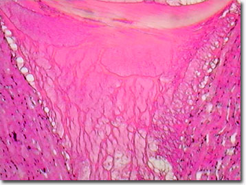
Human Heart Tissue
The mammalian heart is composed primarily of cardiac (or smooth) muscle cells, but includes blood vessels, nerves, and valves. Structurally and functionally, the heart is an efficient, continuously running pump. Arteries, including the aorta (the main artery), carry oxygenated blood away from the heart, and veins return oxygen-depleted blood to the heart.

View a second image of human heart tissue.
From the initiation of development through the moment of death, the heart pumps, requiring tremendous strength, reliability, and resiliency. The average cardiac muscle contracts and relaxes about 80 times per minute. Contractions push the blood through the chambers and into the blood vessels. The innervating neurons regulate the pace of the heart by controlling the muscle contractions. During rest periods such as sleep, the heart pumps more slowly, while during exercise or periods of agitation, the heart pumps more quickly, sending additional oxygenated blood to the body's muscles.
Enclosed in the pericardium, a membranous sac, and protected by the ribcage in front and back, and the diaphragm below, the human heart is characterized by four chambers (two ventricles and two atria) separated by a muscular wall (or septum). Valves connect each atrium to the ventricle below it. Despite the heavy load the heart must bear, the average adult heart is only about the size of a clenched fist and weighs about 285 to 343 grams (10 to 12 ounces) in adult men and 229 to 285 grams (8 to 10 ounces) in adult women.
Contributing Authors
Cynthia D. Kelly, Thomas J. Fellers and Michael W. Davidson - National High Magnetic Field Laboratory, 1800 East Paul Dirac Dr., The Florida State University, Tallahassee, Florida, 32310.
BACK TO THE BRIGHTFIELD IMAGE GALLERY
BACK TO THE DIGITAL IMAGE GALLERIES
Questions or comments? Send us an email.
© 1995-2025 by Michael W. Davidson and The Florida State University. All Rights Reserved. No images, graphics, software, scripts, or applets may be reproduced or used in any manner without permission from the copyright holders. Use of this website means you agree to all of the Legal Terms and Conditions set forth by the owners.
This website is maintained by our
Graphics & Web Programming Team
in collaboration with Optical Microscopy at the
National High Magnetic Field Laboratory.
Last Modification Friday, Nov 13, 2015 at 01:19 PM
Access Count Since September 17, 2002: 10837
Visit the website of our partner in introductory microscopy education:
|
|
