
Brightfield Digital Image Gallery
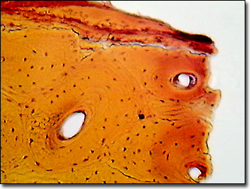
Human Compact Bone
There are two basic structural types of bone in mammals, compact and spongy. Compact bone is very dense and hard on the outside, and makes up most of the bones in the arms and legs of humans.
View a second image of human compact bone.
The structural units of human compact bone are osteons, which are elongated cylinders. Acting as weight-bearing pillars, osteons can withstand high levels of mechanical stress. At the osteon's center is a hollow canal that acts as a central passageway for blood vessels and nerves. A thin membrane, termed the periosteum, surrounds compact bone. The outer layer of this membrane contains blood vessels and nerves that travel into the bone, while the inner layer consists mainly of osteoblasts. These are cells that are constantly renewed in the bone, and which produce and secrete collagen, giving bone a measure of elasticity. In addition, osteoblasts secrete mineral salts formed from calcium and phosphorus, which prevent the bones from breaking easily, as well as rebuilding bone if necessary.
Bones are the major components of skeletal systems in nearly every vertebrate animal. There are 206 bones in the adult human, which make up 14 percent of the total body weight. Bone is composed of both living and nonliving materials. By providing a framework for the body and a surface for the attachment of muscles, the compact bones allow humans to walk upright. Protection for vital organs is provided by other bones, as exemplified by the skull and rib bones, which protect the brain and the heart. Bones also play a major role in human physiology by providing storage for the calcium that is essential for functioning nerve and muscle cells. Additionally, the soft core of bone, the bone marrow, serves as the site for formation of red blood cells, certain white cells, and blood platelets.
Contributing Authors
Cynthia D. Kelly, Thomas J. Fellers and Michael W. Davidson - National High Magnetic Field Laboratory, 1800 East Paul Dirac Dr., The Florida State University, Tallahassee, Florida, 32310.
BACK TO THE BRIGHTFIELD IMAGE GALLERY
BACK TO THE DIGITAL IMAGE GALLERIES
Questions or comments? Send us an email.
© 1995-2025 by Michael W. Davidson and The Florida State University. All Rights Reserved. No images, graphics, software, scripts, or applets may be reproduced or used in any manner without permission from the copyright holders. Use of this website means you agree to all of the Legal Terms and Conditions set forth by the owners.
This website is maintained by our
Graphics & Web Programming Team
in collaboration with Optical Microscopy at the
National High Magnetic Field Laboratory.
Last Modification Friday, Nov 13, 2015 at 02:19 PM
Access Count Since September 17, 2002: 17273
Visit the website of our partner in introductory microscopy education:
|
|