
Brightfield Digital Image Gallery
Frog Striated Muscle
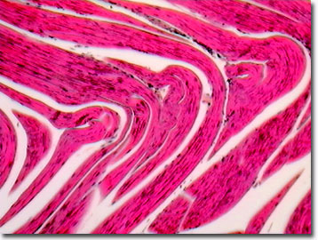
The celebrated jumping frog of Mark Twain's Calaveras County, California would not have been so famous (nor would Mr. Twain) without striated muscles. Striated muscles typical of the rear leg skeletal muscles enable frogs to leap long distances. Composed of narrow and wide elongated fibers, striated muscles appear striped when observed under a microscope.
View a second image of frog striated muscle.
Frogs depend on three types of muscle: smooth, cardiac, and striated. The striated, or skeletal, muscles are bundled together in cords and connect to bones via tendons. Striated muscle cells are among the largest cells in the body of a vertebrate. A favorite of commercial slide preparers, frog striated muscle allows the histology student to view and study the transverse striations, nucleus, collagen tissue, endothelium, and tough surrounding epimysium typical of vertebrate skeletal muscle cells. Physiologists and biophysicists employ frog striated muscle cells as working models for explaining the sliding filament theory and for assessing the mechanical forces and velocities involved with leg (or other skeletal) movement. Lab dissections of the exposed femur, the knee, and the gastrocnemius muscle with attached Achilles tendon allow the researcher to measure response when electrical shocks are applied to contract the muscle.
Contributing Authors
Cynthia D. Kelly, Thomas J. Fellers and Michael W. Davidson - National High Magnetic Field Laboratory, 1800 East Paul Dirac Dr., The Florida State University, Tallahassee, Florida, 32310.
BACK TO THE BRIGHTFIELD IMAGE GALLERY
BACK TO THE DIGITAL IMAGE GALLERIES
Questions or comments? Send us an email.
© 1995-2025 by Michael W. Davidson and The Florida State University. All Rights Reserved. No images, graphics, software, scripts, or applets may be reproduced or used in any manner without permission from the copyright holders. Use of this website means you agree to all of the Legal Terms and Conditions set forth by the owners.
This website is maintained by our
Graphics & Web Programming Team
in collaboration with Optical Microscopy at the
National High Magnetic Field Laboratory.
Last Modification Friday, Nov 13, 2015 at 02:19 PM
Access Count Since September 17, 2002: 19728
Visit the website of our partner in introductory microscopy education:
|
|