
Brightfield Digital Image Gallery
Pig Tooth Enamel Formation
In part because pigs are mammalian omnivores, their teeth can be utilized in studies modeling the development and aging of human teeth. Dental enamel is formed by the epithelial cells of the enamel organ, including the ameloblasts, the cells that produce enamel matrix proteins.
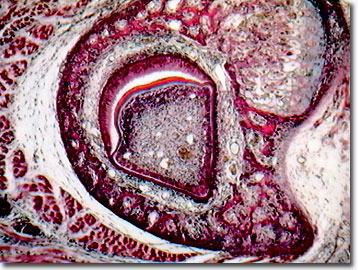
In similarity to humans, pigs display molars, premolars (or bicuspids), canines, and incisors, and, as most mammals, both pigs and humans are diphyodont (develop and erupt two generations of teeth). In the pig's deciduous (baby teeth) complement, there are three incisors, one canine, and three premolars on each side of the bottom jaw for a total of 28 teeth, as compared to humans, which have 20 primary teeth. These 20 grow into a mixed dentition of permanent and primary teeth that ends by the age of 13 years. In their permanent tooth array, pigs exhibit a dentition of three incisors, one canine, four premolars, and three molars on each side of the top and bottom of the mouth, for a total of 44 teeth. Humans, on the other hand, are characterized by a permanent dentition of 32 teeth, 16 upper, 16 lower. Baby pigs undergo the painful process of teething just as that suffered by human babies.
Pig and human teeth are protected by outer coatings of enamel, the hardest tissue in the body. Below the crown line, the root of the tooth is protected by cementum. The bulk of the tooth, below the enamel and cementum, is composed of the dentin, which lines the pulp cavity. Within the central, innermost portion, the pulp performs the formative, sensory, nutritive, and other functions that sustain the life of the tooth. Through the application of tissue culture techniques in the laboratory, enamel protein matrices have been produced from ameloblasts using secreted enamelysin, an active proteolytic enzyme, to process and slowly degrade proteins in secretory stage enamel.
Contributing Authors
Cynthia D. Kelly, Thomas J. Fellers and Michael W. Davidson - National High Magnetic Field Laboratory, 1800 East Paul Dirac Dr., The Florida State University, Tallahassee, Florida, 32310.
BACK TO THE BRIGHTFIELD IMAGE GALLERY
BACK TO THE DIGITAL IMAGE GALLERIES
Questions or comments? Send us an email.
© 1995-2025 by Michael W. Davidson and The Florida State University. All Rights Reserved. No images, graphics, software, scripts, or applets may be reproduced or used in any manner without permission from the copyright holders. Use of this website means you agree to all of the Legal Terms and Conditions set forth by the owners.
This website is maintained by our
Graphics & Web Programming Team
in collaboration with Optical Microscopy at the
National High Magnetic Field Laboratory.
Last Modification Friday, Nov 13, 2015 at 02:19 PM
Access Count Since September 17, 2002: 20183
Visit the website of our partner in introductory microscopy education:
|
|