
Brightfield Digital Image Gallery
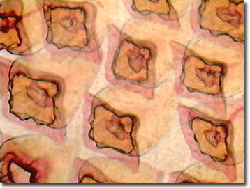
Dogfish Shark Placoid Scales
Dogfish sharks are small sharks belonging to one of three families: the dogfish shark family, Squalidae; the requiem shark family, Carcharhinidae; and the cat shark family, Scyliorhinidae. The best-known species are the spiny dogfish of the dogfish shark family, and the smooth dogfish of the requiem shark family.
Dogfish sharks make up one subfamily of the dogfish shark family, and are characterized by a hard spine at the base of each of the two dorsal fins. Other species of the dogfish shark family that are called dogfish include the Atlantic black dogfish and the green dogfish, found in deep waters of the Gulf of Mexico. The green dogfish, along with several other members of the dogfish shark family, is luminescent.
Scales are small plates that form part of the skin of certain animals. They provide protection from the environment and from predators. Sharks have placoid scales, bony, spiny projections with an enamel-like covering. These scales have the same structure as the fish's teeth, and are also referred to as dermal denticles ( dermal=skin, denticle=teeth). These denticles are slanted toward the tail of the shark and help direct the flow of water around the shark's body, reducing friction so it can swim with less effort. Stroking a shark from head to tail, its skin feels smooth to the touch, but stroking it from tail to head, the skin feels as rough as sandpaper. Shark's skin has, in fact, been used as sandpaper in some countries for many centuries. A shark can also inflict wounds on potential prey by breaking the creature's skin with its scales. Like teeth, the shape of the scales varies among shark species and can be used to identify them.
Contributing Authors
Cynthia D. Kelly, Thomas J. Fellers and Michael W. Davidson - National High Magnetic Field Laboratory, 1800 East Paul Dirac Dr., The Florida State University, Tallahassee, Florida, 32310.
BACK TO THE BRIGHTFIELD IMAGE GALLERY
BACK TO THE DIGITAL IMAGE GALLERIES
Questions or comments? Send us an email.
© 1995-2025 by Michael W. Davidson and The Florida State University. All Rights Reserved. No images, graphics, software, scripts, or applets may be reproduced or used in any manner without permission from the copyright holders. Use of this website means you agree to all of the Legal Terms and Conditions set forth by the owners.
This website is maintained by our
Graphics & Web Programming Team
in collaboration with Optical Microscopy at the
National High Magnetic Field Laboratory.
Last Modification Friday, Nov 13, 2015 at 02:19 PM
Access Count Since September 17, 2002: 17759
Visit the website of our partner in introductory microscopy education:
|
|