
Brightfield Digital Image Gallery
Dictydium cancellatum
The digital image presented below illustrates the slime mold fungus Dictydium cancellatum under brightfield illumination, captured at high magnification (at the top end of the zoom range) with the MIC-D inverted microscope.
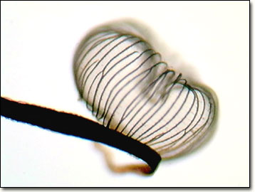
Mycologists commonly study slime molds because their fruiting body parts have many similarities in appearance to common fungi. These tiny creatures are usually found in rotted logs and decaying plant matter, where there is an abundant supply of moisture and bacteria. Slime molds produce spores, which germinate to form myxamoebae, a flagellated swarm cell that eventually fuses to generate the plasmodium intermediate, and ultimately, the fruiting body.
Contributing Authors
Cynthia D. Kelly, Thomas J. Fellers and Michael W. Davidson - National High Magnetic Field Laboratory, 1800 East Paul Dirac Dr., The Florida State University, Tallahassee, Florida, 32310.
BACK TO THE BRIGHTFIELD IMAGE GALLERY
BACK TO THE DIGITAL IMAGE GALLERIES
Questions or comments? Send us an email.
© 1995-2025 by Michael W. Davidson and The Florida State University. All Rights Reserved. No images, graphics, software, scripts, or applets may be reproduced or used in any manner without permission from the copyright holders. Use of this website means you agree to all of the Legal Terms and Conditions set forth by the owners.
This website is maintained by our
Graphics & Web Programming Team
in collaboration with Optical Microscopy at the
National High Magnetic Field Laboratory.
Last Modification Friday, Nov 13, 2015 at 02:19 PM
Access Count Since September 17, 2002: 16633
Visit the website of our partner in introductory microscopy education:
|
|