
Brightfield Digital Image Gallery
Fat-Stained Adipose Tissue
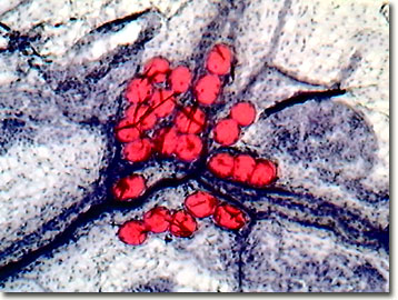
Adipose tissue is a specialized connective tissue that serves as a main storage site for triglycerides (fat), and is found in two forms, brown and white, in mammals. The digital image presented below was captured with the MIC-D and reveals fat globules in a thin section of adipose tissue stained with Sudan IV.
White adipose tissue serves three main functions, but its primary one is to serve as a storage location for energy, in the form of long-chain fatty acids and triglycerides. The tissue also serves secondary duties as a mechanical cushion to protect against impact, and as an insulator to keep the host warm. Located directly beneath the skin, subcutaneous adipose tissue is an especially important heat insulator that has a conductance value approximately one-third that of other tissues.
Deriving its color character from rich vascularization and densely packed mitochondria, brown adipose tissue is found in a variety of locations, depending upon the species and age of the animal. This tissue is metabolically less active in non-hibernating animals, although exposure to cold weather can activate the tissue. In adult mammals, the majority of bulk adipose tissue is composed of a loose association of lipid-filled cells termed adipocytes, which are connected together in a framework of collagen fibers.
Contributing Authors
Cynthia D. Kelly, Thomas J. Fellers and Michael W. Davidson - National High Magnetic Field Laboratory, 1800 East Paul Dirac Dr., The Florida State University, Tallahassee, Florida, 32310.
BACK TO THE BRIGHTFIELD IMAGE GALLERY
BACK TO THE DIGITAL IMAGE GALLERIES
Questions or comments? Send us an email.
© 1995-2025 by Michael W. Davidson and The Florida State University. All Rights Reserved. No images, graphics, software, scripts, or applets may be reproduced or used in any manner without permission from the copyright holders. Use of this website means you agree to all of the Legal Terms and Conditions set forth by the owners.
This website is maintained by our
Graphics & Web Programming Team
in collaboration with Optical Microscopy at the
National High Magnetic Field Laboratory.
Last Modification Friday, Nov 13, 2015 at 02:19 PM
Access Count Since September 17, 2002: 19695
Visit the website of our partner in introductory microscopy education:
|
|