
Polarized Light Microscopy
Adapting the Intel Play QX3 microscope for use with plane-polarized light is one of the simplest and most useful configurations available for this amazing microscope. Polarized light microscopy allows the student to explore the birefringent properties of common household items such as sugar, epsom salts, Kool Aid, vitamins, aspirin, and moth balls.
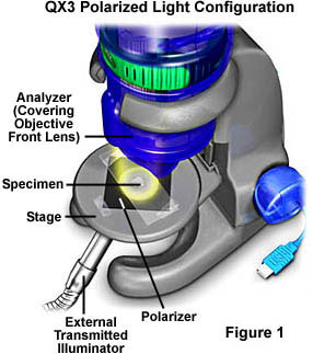
The basic setup for transmitted polarized light using the QX3 microscope is illustrated in Figure 1. A polarizer is added to the optical pathway somewhere beneath the specimen, either on the stage or within the mixing chamber (Figure 4). Another polarizer, termed the analyzer is placed over the front lens of the objective in the body of the microscope (Figure 3).
An external tungsten-halide light source with a fiber optics light guide is used to provide auxiliary illumination (Figure 1), because polarizers can block up to 90 percent of the light passing through them. A quarter-inch hole is drilled at the base of the substage mixing chamber to allow light from the illuminator to enter. The mixing chamber will assist in diffusing the light and minimize any problems with bright spots in the image.
Light passing through a polarizer is polarized in a single vibrational plane by a polymer film that is highly oriented within the polarizer. Polarizing films are not very expensive and can be readily purchased from many science supply houses such as Edmund Scientific, Fisher, Carolina Biological Supply, and Ward's. Edmund Scientific markets linear polarizing sheet film in a sample pack containing two 2" x 2" squares for $5.95, and they also offer a Polarizer Experimenter's Kit containing polarizers, quarter- and half-wave retardation plates, and a blue blocking polarizer for $23.00 plus shipping.
| Interactive Java Tutorial | |||||||||||
|
|||||||||||
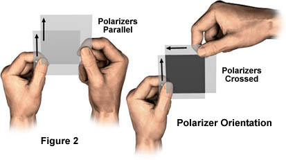
The polarizers must be installed with a specific orientation in the microscope to ensure their polarization vectors are crossed (perpendicular). Two sets of polarizers in different orientations are illustrated in Figure 2. Black arrows on the polarizers indicate the direction of the polarization vector. The pair of hands to the left in Figure 2 are holding two polarizers that have the polarization vectors aligned parallel and the pair of hands on the right are holding the polarizers with their vectors perpendicular (crossed polarizers). It is the arrangment of crossed polarizers on the right-hand side of Figure 2 that is useful for polarized light microscopy.
When two polarizers are held together with their polarization vectors parallel to each other as shown in Figure 2, then light polarized and passed by the first filter will be in the correct orientation to pass through the second. The polarizers will appear transparent when held before a strong light source. However, when the polarizers are held together with their polarization vectors perpendicular, the second polarizer will no longer pass light polarized by the first. In this case, holding the crossed polarizers before a strong light source will demonstrate that no light passes through.
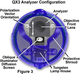
After the vibrational planes have been identified for the polarizers, the next job is to attach them in a fixed position on the microscope. Carefully cut one of the polarizers into the shape depicted in Figure 3 for placement in front of the objective lenses. The white arrow in Figure 3 indicates the correct direction of the vibrational plane for the polarizer placed in the microscope body. This polarizer, termed the analyzer and having a specific orientation by convention, is placed with the vibrational plane in a North-South (front to back) direction. Secure the analyzer tightly on the clear dust cover protecting the objective's lens.
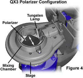
The next step is to place a polarizer either on top of the diffusion screen in the stage or beneath the screen in the mixing chamber. Illustrated in Figure 4 is a polarizer secured in the mixing chamber and showing the orientation of the vibrational plane of the polarizer (white double-headed arrow). This polarizer is the one responsible for the initial polarization of white light before it illuminates the sample. Also by convention, this polarizer is oriented in an East-West (left to right) direction with respect to the microscope body. It is often more convenient, especially when frequently interchanging illumination modes, to simply place the polarizer on the stage underneath the specimen (Figure 1). When correctly oriented, the Live View window should be very dark or black when the microscope is illuminated but no specimen is on the stage.
Once the polarizers are installed, the microscope is ready to image birefringent specimens. Candidates for polarized light microscopy include any number of common chemicals found around the home or in the supermarket or drug stores. Many vitamins can be recrystallized to form colorful patterns when imaged between crossed polarizers. Likewise, sugar can be dissolved in water and the solution sandwiched between a microscope slide and coverslip, then allowed to slowly evaporate. For more suggestions, visit our section on Specimen preparation for polarized light microscopy.
| Interactive Java Tutorial | |||||||||||
|
|||||||||||
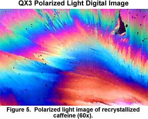
The photomicrograph depicted in Figure 5 illustrates birefringent crystallites of the caffeine, a naturally occuring chemical found in coffee and added to many soft drinks. The specimen was prepared through the melt-recrystallized technique in which a few micrograms of pure crystalline power are placed on a microscope slide and melted with a Bunsen burner. After melting, the slide is placed on the counter top and the crystals are allowed to slowly form from the melt. After the melted caffeine has completely recrystallized and the slide has cooled to room temperature, it is placed on the microscope stage and digitally imaged under polarized light. Vitamin C, aspirin, and acetamidophen (Tylenol) are also good candidates for quick melt-recrystallization experiments in polarized light.
Only those specimens that display birefringence will be visible under polarized illumination. This includes most crystals such as sugar, drugs, and vitamins, some spices and herbs, rock thin sections, bone sections, feathers, butterfly wings, fish scales, and a wide spectrum of other specimens. Part of the fun of polarized light microscopy is to determine which specimens are birefringent. For instance, table salt is one of the most readily available crystals, but the chemical nature of the salt crystal makes it optically isotropic, or invisible in polarized light. Hairs and fibers are often birefringent and make excellent candidates for polarized light microscopy, but materials like wood, metals, and opaque plastics are not. A careful examination of specimens immediately available will help initiate the course towards successful polarized light microscopy.
You can learn more about birefringence and polarized light, along with a number of other topics in the physics of light and color and optical microscopy in our Molecular Expressions Microscopy Primer website.
BACK TO INTEL QX3 SPECIALIZED TECHNIQUES
Questions or comments? Send us an email.
© 1995-2025 by Michael W. Davidson and The Florida State University. All Rights Reserved. No images, graphics, software, scripts, or applets may be reproduced or used in any manner without permission from the copyright holders. Use of this website means you agree to all of the Legal Terms and Conditions set forth by the owners.
This website is maintained by our
Graphics & Web Programming Team
in collaboration with Optical Microscopy at the
National High Magnetic Field Laboratory.
The QX3 microscope design is copyrighted © 2025 by Mattel, Inc. Intel® Play™ is a registered trademark of the Intel Corporation. These companies reserve all of their rights and privileges under copyright law. The material contained in this website is solely the opinion of the authors and is intended for eduational use only.
Last Modification Friday, Nov 13, 2015 at 02:18 PM
Access Count Since December 24, 1999: 55693
Visit the official Intel Play website:
![]()