
Advanced Condenser Systems: Abbe Condensers
Silkworm Larva Spiracle & Trachea
The images below compare performance of the Intel Play QX3 Computer Microscope with and without the aid of an organized cone of illumination from an Abbe substage condenser containing an aperture diaphragm. These photomicrographs are unretouched and were captured with the QX3 interactive software.
Unstained Silkworm Larva
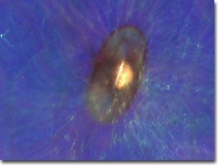
(Blue Central and Red/Yellow Annular Gels)

with Abbe Condenser
(Blue Central and Red/Yellow Annular Gels)
"Silkworm" is the common name for silk-producing larvae of any of several species of moths. The most celebrated is the common, domesticated silkworm that was originally cultivated by the Chinese 5,000 years ago. These larvae feed on leaves of white mulberry, Osage orange, or lettuce, although artificial diets have been developed for modern commercial operations. After six weeks, the silkworm stops eating and spins its cocoon. The larvae possess a pair of specially modified salivary glands, called silk glands. These glands secrete a clear, viscous fluid that is forced through openings, called spinnerets on the mouthparts of the larva. The fluid hardens as it comes into contact with air to become the silk thread. The diameter of the spinneret determines the thickness of the silk thread produced. Each cocoon of yields a strand of silk about 1,000 yards (900 meters) long.
Silkworm moths live only in captivity at present and are grown in commercial operations around the world. All wild populations are extinct, although presumably there are still related species in Asia. Silkworms have been domesticated so long that they have lost the ability to fly and can no longer survive independently in nature.
Semi-transparent specimens are difficult to image using unaided brightfield optical microscopy. The images above were recorded using the Intel Play QX3 microscope in transmitted brightfield mode with the assistance of Rheinberg illumination. Dyed acetate gel filters were strategically placed below the aperture diaphragm of an aftermarket Abbe-style condenser to generate the Rheinberg effect. A 15-millimeter central filter was surrounded by a larger annular filter to combine the effects of oblique specimen illumination by light filtered through the annular filter superimposed over a background of light filtered through the central filter.
BACK TO INTEL QX3 ADVANCED RHEINBERG GALLERY
Questions or comments? Send us an email.
© 1995-2025 by Michael W. Davidson and The Florida State University. All Rights Reserved. No images, graphics, software, scripts, or applets may be reproduced or used in any manner without permission from the copyright holders. Use of this website means you agree to all of the Legal Terms and Conditions set forth by the owners.
This website is maintained by our
Graphics & Web Programming Team
in collaboration with Optical Microscopy at the
National High Magnetic Field Laboratory.
The QX3 microscope design is copyrighted © 2025 by Mattel, Inc. Intel® Play™ is a registered trademark of the Intel Corporation. These companies reserve all of their rights and privileges under copyright law. The material contained in this website is solely the opinion of the authors and is intended for eduational use only.
Last Modification Friday, Nov 13, 2015 at 02:19 PM
Access Count Since April 25, 2000: 17021
Visit the official Intel Play website:
![]()