
Anatomy of the MIC-D Digital Microscope
Olympus has thrown the doors open to a new era in optical microscopy education with the introduction of the MIC-D inverted digital microscope. Designed specifically for a wide spectrum of applications ranging from basic classroom instruction to more advanced laboratory analysis, this versatile microscope features a palette of contrast enhancing techniques that rival many research-level instruments.
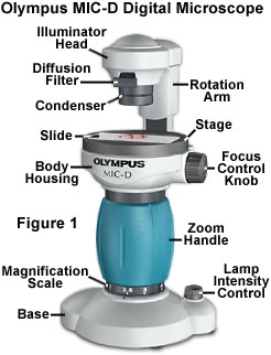
The MIC-D digital microscope incorporates cutting-edge light-emitting diode (LED) solid-state illumination technology in a swivel lamphouse (rotation arm) that also contains a diffusion screen and condenser lens (see Figure 1). This combination of lighting elements enables the microscopist to illuminate the specimen from a variety of off-axis angles and different numerical apertures. Illumination modes include traditional brightfield, oblique, darkfield, and reflected light. The Olympus MIC-D digital microscope should set a standard for future microscopes in the coming years by affording a variety of contrast-enhancing techniques that are unparalleled in competing microscopes of this class.
Anatomical Overview - The Olympus MIC-D digital microscope represents a unique design that incorporates a CMOS electronic digital imaging sensor as a substitute for the traditional eyepieces or oculars found in a majority of microscopes. Coupled to an inverted illumination system that broadcasts light from above the specimen (similar to a tissue culture microscope), this digital microscope also features a translatable lamphouse and condenser unit that can be rotated over a 135-degree angle. Such versatility allows the operator to project light onto the specimen from an almost limitless combination of oblique or reflected illumination angles.
Electrical and Image Sensor Control System - Because the Olympus MIC-D digital microscope operates with the assistance of electrical current derived from a host computer, it does not require batteries or an external alternating current power supply. The microscope has a rated operating potential of 5.0 volts at a nominal current of 0.4 amperes, and receives power through a Universal Serial Bus (USB) interface from the local host computer. The power supplied by the host computer serves to provide current to the microscope illumination and image sensor components, which are the primary electrical (and electronic) systems that control luminous flux and image capture with the microscope.
Brightfield Illumination - Transmitted brightfield illumination is one of the most commonly utilized observation modes in optical microscopy, and is ideal for fixed, stained specimens or other types of samples having high natural absorption of visible light. Collectively, specimens imaged with brightfield illumination are termed amplitude objects because the amplitude or intensity of the illuminating wavefronts is reduced when light passes through the specimen. When the illuminator rotation arm of the Olympus MID-D digital microscope is placed in the vertical position, and the illuminator housing is coaxial with the optical train, the microscope can operate in transmitted brightfield mode.
Oblique Illumination - In oblique illumination, direct light from the condenser light cone is restricted to a single azimuth, striking the specimen from only one direction rather than bathing it with an even distribution of light. The net effect is to reveal details in pseudo-relief to produce a shadowed effect on the side opposite the light source and bright specimen highlights on the side nearest the illuminator. In order to achieve oblique or off-axis illumination with the MIC-D digital microscope, the illuminator head is shifted by moving the rotation arm a few degrees away from the upright (brightfield) position.
Darkfield Illumination - In traditional optical microscopy, darkfield illumination requires blocking the central light that ordinarily passes through and around (surrounding) the specimen, allowing only oblique rays from every azimuth to "strike" the specimen mounted on a microscope slide. When the rotation arm of the MIC-D digital microscope is positioned at a distance greater than 15 degrees from the central (optical) axis, this instrument is capable of imaging specimens by a mechanism that produces results similar to those observed in true darkfield illumination.
Polarized Illumination - The polarized light microscope is designed to observe and record images of specimens that are visible primarily due to their optically anisotropic character. These specimens have polarizable intramolecular bonds that interact with polarized light in a direction-sensitive manner to produce phase retardations that are monitored through interference-dependent alterations in amplitude in the image plane. The MIC-D microscope can be equipped with a polarizer and analyzer combination in order to view and capture digital images from birefringent anisotropic specimens.
Reflected Illumination - In general terms, the world of optical microscopy can be largely divided into two main categories: transmitted light microscopy and reflected light microscopy. Transmitted light microscopy is utilized for specimens that are relatively thin and semi-transparent, enabling a significant amount of light to pass through. In contrast, reflected light microscopy, often termed incident light or metallurgical microscopy, is reserved for those specimens that remain opaque even when ground to a thickness of 30 micrometers or less. The MIC-D digital microscope can easily be configured to capture digital images in reflected light illumination.
Interactive Java Tutorials
MIC-D Microscope Focus Mechanism - The Olympus MIC-D digital microscope is equipped with a single knob, located on the upper microscope body beside the gliding stage, which enables the operator to focus specimens. The microscope focus mechanism is simple in design, yet affords an easy method to quickly bring specimen details into fine focus throughout the microscope zoom magnification range. This tutorial describes lens proximity variations during the focusing operation and illustrates how the components work together to achieve focus.
Rotation Arm Range of Motion - In order to obtain a variety of illumination modes, the Olympus MIC-D digital microscope is equipped with a rotation arm that travels through a range of 135 degrees. Using the various positions available with the rotation arm, the microscope can be configured for brightfield, oblique, darkfield, and reflected light observation. This tutorial explores the range of motion available with the MIC-D rotation arm.
Image-Forming and Aperture Light Pathways - The Olympus MIC-D digital microscope is equipped with a zoom lens system that operates in conjunction with the objective lens elements to form an image of the specimen on the surface of the CMOS image sensor. This interactive tutorial explores both the image forming and aperture light ray pathways through the microscope zoom optical body and how they are affected by changing the position of individual lens elements.
Off-Axis Illumination Transitions - The true versatility of the unique MIC-D digital microscope design becomes apparent with oblique and darkfield illumination techniques made possible by off-axis translation of the illuminator head and condenser assembly. This feature enables the microscopist to enhance contrast in specimens that would otherwise remain invisible (or nearly so) in brightfield illumination. The interactive tutorial explores illumination transitions, starting at brightfield, moving through the varying degrees of oblique, and finally reaching darkfield.
MIC-D Zoom Lens System - The MIC-D digital microscope is equipped with a zoom lens system that aids the objective in forming a magnified image of the specimen. Two individual lens elements combine to form the zoom system, and these are translated simultaneously, guided by a track system, within the microscope zoom handle to alter the microscope magnification factor. This interactive tutorial explores the position of the zoom lens elements at various magnification factors.
Contributing Authors
Thomas J. Fellers and Michael W. Davidson - National High Magnetic Field Laboratory, 1800 East Paul Dirac Dr., The Florida State University, Tallahassee, Florida, 32310.
BACK TO THE OLYMPUS MIC-D DIGITAL MICROSCOPE
Questions or comments? Send us an email.
© 1995-2022 by Michael W. Davidson and The Florida State University. All Rights Reserved. No images, graphics, software, scripts, or applets may be reproduced or used in any manner without permission from the copyright holders. Use of this website means you agree to all of the Legal Terms and Conditions set forth by the owners.
This website is maintained by our
Graphics & Web Programming Team
in collaboration with Optical Microscopy at the
National High Magnetic Field Laboratory.
Last Modification Friday, Nov 13, 2015 at 02:19 PM
Access Count Since September 17, 2002: 47209
Visit the website of our partner in introductory microscopy education:
|
|