
Interactive Tutorial
MIC-D Microscope Focus Mechanism
The Olympus MIC-D digital microscope is equipped with a single knob, located on the upper microscope body beside the gliding stage, which enables the operator to focus specimens. The microscope focus mechanism is simple in design, yet affords an easy method to quickly bring specimen details into fine focus throughout the microscope zoom magnification range. This tutorial describes lens proximity variations during the focusing operation and illustrates how the components work together to achieve focus.
The tutorial initializes with a graphical depiction of the MIC-D upper microscope body cutaway to reveal the objective lens system. To operate the tutorial, use the Adjust Focus slider to translate the objective front lens element with respect to the second lens. As the slider is moved, the Focus Knob rotates to demonstrate how the focus mechanism operates.
Removing the MIC-D digital microscope stage exposes the movable objective front lens element and the Focus Control Mechanism, which are illustrated in Figure 1. Similar to the condenser lens, the objective front lens is bi-convex with surfaces having dissimilar radii. The surface having a larger radius (the "flatter" side of the lens) is almost flat and faces the specimen, while the surface with a smaller radius (greater curvature) faces the second objective lens element. This lens element is mounted in a machined black-anodized aluminum housing that has an extended annular ring with a groove cut into the center. A pin on the focus control mechanism fits into the groove and is used to translate the lens mount upward and downward when the focus knob is rotated.
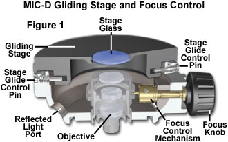
The focus knob is mounted on a partially threaded shaft that is guided by a machined boss cast into the microscope upper body unit. A 10-millimeter length of brass rod is drilled and tapped to thread onto the focus knob shaft. On the end of the rod facing away from the knob is a small pin that fits into the lens mount groove. When the focus knob is rotated, the pin scribes an orbital path that translates the objective front lens to enable the operator to focus the specimen.
A second objective bi-concave lens element (see the MIC-D Objective Assembly illustrated in Figure 2) is mounted in a seat containing a 9-millimeter aperture machined into a boss cast in the center of the microscope optical axis. This lens element is fixed into position and serves to focus light from the front objective lens element into the zoom optical system. The aluminum mount holding the first objective lens glides over the cast aluminum boss (containing the second lens), which holds the upper objective assembly together.
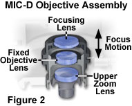
Coupled to the two upper lens elements to form a working objective is the first lens in the zoom optical system, which serves as the final objective lens, and is seated in a counter-weighted translatable mount that glides up and down the body tube of the microscope as the magnification is varied. A 9-millimeter diameter aperture diaphragm built into the mounting seat of this lens element acts as a primary conjugate plane for the illuminating (aperture) light flux passing through the microscope. The second lens of the zoom system performs the function of a relay lens to assist the projection lens in forming a focused image of the specimen on the surface of the CMOS image sensor.
Contributing Authors
Matthew J. Parry-Hill and Michael W. Davidson - National High Magnetic Field Laboratory, 1800 East Paul Dirac Dr., The Florida State University, Tallahassee, Florida, 32310.
BACK TO MIC-D MICROSCOPE ANATOMY
BACK TO THE OLYMPUS MIC-D DIGITAL MICROSCOPE
Questions or comments? Send us an email.
© 1995-2022 by Michael W. Davidson and The Florida State University. All Rights Reserved. No images, graphics, software, scripts, or applets may be reproduced or used in any manner without permission from the copyright holders. Use of this website means you agree to all of the Legal Terms and Conditions set forth by the owners.
This website is maintained by our
Graphics & Web Programming Team
in collaboration with Optical Microscopy at the
National High Magnetic Field Laboratory.
Last Modification Tuesday, Sep 11, 2018 at 03:44 PM
Access Count Since September 17, 2002: 25479
Visit the website of our partner in introductory microscopy education:
|
|