Fluorescence Digital Image Gallery
Human Bone Osteosarcoma Cells (U-2 OS)
|
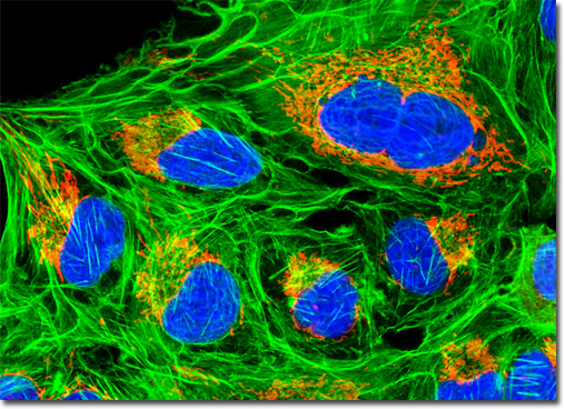
Traditionally, treatment for osteosarcoma consisted of the amputation of the affected limb. However, medical advances over the last several decades have made amputation much less common. Today surgeons often remove the tumor and a small area of healthy tissue surrounding it without finding it necessary to remove the entire bone. The bone tissue that is lost is typically replaced with either an artificial implant or bone material taken from another part of the body through a process known as a bone graft. Chemotherapy or radiation therapy may also be included in osteosarcoma treatment, the former primarily being utilized to destroy any remaining malignant cells in the body following surgery and the latter sometimes being used locally in cases where surgical removal of the tumor is not feasible. The culture of human osteosarcoma cells featured in the digital image above was transfected with a pDsRed-Mitochondria plasmid subcellular localization vector, thus localizing a red fluorescent protein tag to the intracellular mitochondrial network. After isolation of stable transfectants, a healthy culture was fixed and subsequently labeled with DAPI and Alexa Fluor 488 conjugated to phalloidin, targeting DNA and F-actin, respectively. Images were recorded in grayscale with a QImaging Retiga Fast-EXi camera system coupled to an Olympus BX-51 microscope equipped with bandpass emission fluorescence filter optical blocks provided by Omega Optical. During the processing stage, individual image channels were pseudocolored with RGB values corresponding to each of the fluorophore emission spectral profiles. |
© 1995-2025 by Michael W. Davidson and The Florida State University. All Rights Reserved. No images, graphics, software, scripts, or applets may be reproduced or used in any manner without permission from the copyright holders. Use of this website means you agree to all of the Legal Terms and Conditions set forth by the owners.
This website is maintained by our
|
