Fluorescence Digital Image Gallery
Human Bone Osteosarcoma Cells (U-2 OS)
|
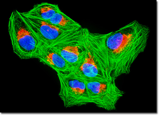
In dogs, osteosarcoma is often first suspected when the animal begins to limp or becomes lame. Similar to many other animal diseases, osteosarcoma is frequently left untreated in pets due to expense, with owners often choosing to have the animal euthanized when their pain seems to hinder quality of life. However, due to the recent availability of pet insurance, it is becoming increasingly common for animals to receive osteosarcoma treatments comparable to those received by humans, including surgery, radiotherapy, and chemotherapy. For dogs, however, complete amputation of the problematic limb is more common than it currently is for people. Most owners observe significant improvement in their pet following such surgery, but because the cancer cells have generally invaded other areas before diagnosis is made, amputation alone does not usually extend the life of the animal, though it does eliminate much of the pain experienced. A dog that is not given any treatment and one that is only provided with surgery do not generally live longer than one or two months following diagnosis. When other therapies are used in conjunction with surgery, the one-year survival rate is about 50 percent. In what has now become a traditional fluorescence staining cocktail, the culture of osteosarcoma cells appearing in the digital image above was stained with MitoTracker Red CMXRos, Alexa Fluor 488 conjugated to phalloidin, and DAPI, in order to target the mitochondrial network, filamentous actin, and nuclear DNA, respectively. Images were recorded in grayscale with a QImaging Retiga Fast-EXi camera system coupled to an Olympus BX-51 microscope equipped with bandpass emission fluorescence filter optical blocks provided by Omega Optical. During the processing stage, individual image channels were pseudocolored with RGB values corresponding to each of the fluorophore emission spectral profiles. |
© 1995-2025 by Michael W. Davidson and The Florida State University. All Rights Reserved. No images, graphics, software, scripts, or applets may be reproduced or used in any manner without permission from the copyright holders. Use of this website means you agree to all of the Legal Terms and Conditions set forth by the owners.
This website is maintained by our
|
