Fluorescence Digital Image Gallery
Human Bone Osteosarcoma Cells (U-2 OS)
|
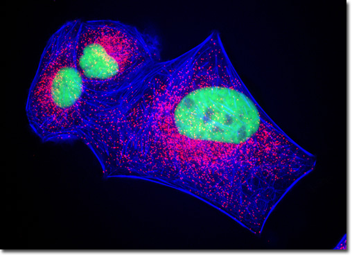
Alexa Fluor dyes are a collection of high-quality fluorescent probes that were designed to correlate with the principal output wavelengths of common excitation sources. The spectra of the dyes are insensitive to pH over a broad range and well differentiated from one another, so that multiple members of the Alexa Fluor line may be utilized simultaneously for multicolor detection experiments. The blue-fluorescent member of the line is Alexa Fluor 350, a sulfonated coumarin derivative. Alexa Fluor 350 protein conjugates are excited optimally at 346 nanometers and emit fluorescence at approximately 442 nanometers, a wavelength somewhat shorter than that emitted by AMCA or AMCA-X conjugates (448 nanometers), which reduces spectral overlap with the popular dye fluorescein. In a double immunofluorescence labeling experiment, the culture of U-2 OS cells featured in the digital image above was treated with a cocktail of mouse anti-histones (pan) and rabbit anti-PMP 70 (peroxisomal membrane protein) primary antibodies. The target proteins were subsequently visualized with goat anti-mouse and anti-rabbit secondary antibodies conjugated to Cy2 and Rhodamine Red, respectively. The filamentous actin cytoskeletal network was counterstained with Alexa Fluor 350 conjugated to phalloidin. Images were recorded in grayscale with a QImaging Retiga Fast-EXi camera system coupled to an Olympus BX-51 microscope equipped with bandpass emission fluorescence filter optical blocks provided by Omega Optical. During the processing stage, individual image channels were pseudocolored with RGB values corresponding to each of the fluorophore emission spectral profiles. |
© 1995-2025 by Michael W. Davidson and The Florida State University. All Rights Reserved. No images, graphics, software, scripts, or applets may be reproduced or used in any manner without permission from the copyright holders. Use of this website means you agree to all of the Legal Terms and Conditions set forth by the owners.
This website is maintained by our
|