Fluorescence Digital Image Gallery
Human Bone Osteosarcoma Cells (U-2 OS)
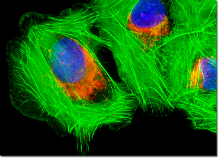
|
As treatment for cancer has improved in recent years, so have the chances of recovery from osteosarcoma. In cases where the disease is diagnosed early in its development, five-year survival rates are as high as 60 to 80 percent. Unfortunately, however, due to the rapid advancement of osteosarcoma, approximately one in every five children diagnosed with the disease are found to already be in an advanced stage, in which cancer cells have begun invading other regions of the body. The five-year survival rate in such cases is usually approximated at less than 10 percent. Also, patients that do survive may be plagued with various side effects and long-term risks from the treatment they received. For instance, because exposure to radiation increases the risk for DNA mutations, patients who have undergone radiation therapy as part of their treatment for osteosarcoma are at increased risk for the development of other cancers later in life. The mitochondria present in the culture of U-2 OS human cancer cells featured in the digital image above were targeted with MitoTracker Red CMXRos, a derivative of X-rosamine. In addition, the culture was labeled for F-actin and nuclear DNA with Alexa Fluor 488 conjugated to phalloidin and DAPI, respectively. Images were recorded in grayscale with a QImaging Retiga Fast-EXi camera system coupled to an Olympus BX-51 microscope equipped with bandpass emission fluorescence filter optical blocks provided by Omega Optical. During the processing stage, individual image channels were pseudocolored with RGB values corresponding to each of the fluorophore emission spectral profiles. |
© 1995-2025 by Michael W. Davidson and The Florida State University. All Rights Reserved. No images, graphics, software, scripts, or applets may be reproduced or used in any manner without permission from the copyright holders. Use of this website means you agree to all of the Legal Terms and Conditions set forth by the owners.
This website is maintained by our
|