Fluorescence Digital Image Gallery
Human Bone Osteosarcoma Cells (U-2 OS)
|
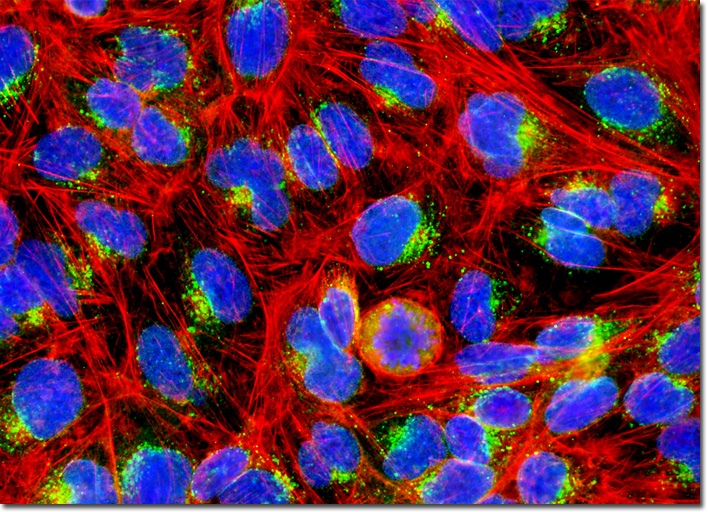
Diagnosis of osteosarcoma may include several different kinds of procedures. Typically, a physical examination takes place first in order to identify any swollen or tender areas of the body. Subsequently, the bone in question may be X-rayed in order to observe any structural changes that are characteristic of osteosarcoma. Although generally signs of a tumor may be detected in this way, a bone biopsy is usually carried out in order to authenticate the diagnosis. In some cases, a computed tomography (CT) scan or magnetic resonance imaging (MRI) are also used to provide a more detailed depiction of the state of the disease, readily revealing whether or not the cancer has spread to surrounding tissues or metastasized to more distant parts of the body. Such tests are also commonly performed after treatment has begun as a way to gauge the extent of the success of the therapy and whether or not the disease is continuing to invade new areas. The adherent culture of human osteosarcoma (U-2 OS) cells illustrated in the digital image above was stained with Oregon Green 488 conjugated to wheat germ agglutinin, a plant-derived lectin that targets the Golgi apparatus, as well as Alexa Fluor 568 conjugated to phalloidin for cytoskeletal actin. Nuclei were labeled with the ultraviolet-absorbing probe DAPI. Images were recorded in grayscale with a QImaging Retiga Fast-EXi camera system coupled to an Olympus BX-51 microscope equipped with bandpass emission fluorescence filter optical blocks provided by Omega Optical. During the processing stage, individual image channels were pseudocolored with RGB values corresponding to each of the fluorophore emission spectral profiles. |
© 1995-2025 by Michael W. Davidson and The Florida State University. All Rights Reserved. No images, graphics, software, scripts, or applets may be reproduced or used in any manner without permission from the copyright holders. Use of this website means you agree to all of the Legal Terms and Conditions set forth by the owners.
This website is maintained by our
|