Fluorescence Digital Image Gallery
Human Bone Osteosarcoma Cells (U-2 OS)
|
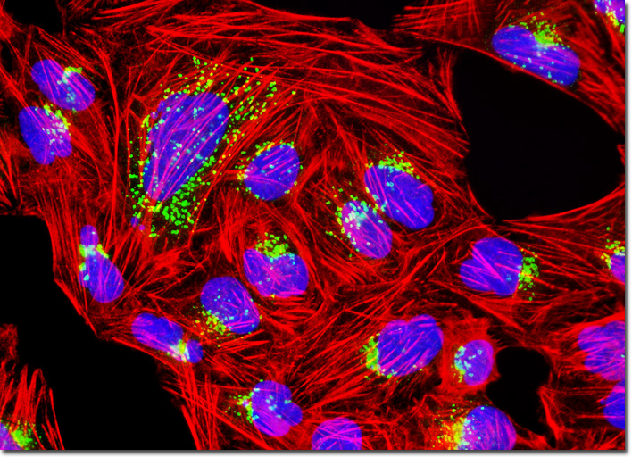
Although survival rates for patients with osteosarcoma are increasing, researchers hope to see even greater improvement in the future. New chemotherapy drugs are continually being developed and may eventually make the eradication of cancer cells a simpler process. Especially beneficial would be the discovery of drugs that solely target tumorous tissue, rather than all rapidly dividing cells. Another promising area for potential osteosarcoma treatments involves new information that is being discovered about the role certain growth factors have in the development of this type of bone cancer. Current studies and research efforts in the field may eventually be utilized to produce new drugs that could slow the generation of these growth factors as way to treat osteosarcoma. The culture of human osteosarcoma (U-2 OS) cells presented in the digital image above was stained with Alexa Fluor 594 conjugated to phalloidin and Hoechst 33342, targeting the cytoskeletal filamentous actin network and nuclear DNA, respectively. The cells were also immunofluorescently labeled with primary anti-giantin rabbit monoclonal antibodies followed by goat anti-rabbit secondary antibodies conjugated to Alexa Fluor 488 in order to target the Golgi apparatus. Images were recorded in grayscale with a QImaging Retiga Fast-EXi camera system coupled to an Olympus BX-51 microscope equipped with bandpass emission fluorescence filter optical blocks provided by Omega Optical. During the processing stage, individual image channels were pseudocolored with RGB values corresponding to each of the fluorophore emission spectral profiles. |
© 1995-2025 by Michael W. Davidson and The Florida State University. All Rights Reserved. No images, graphics, software, scripts, or applets may be reproduced or used in any manner without permission from the copyright holders. Use of this website means you agree to all of the Legal Terms and Conditions set forth by the owners.
This website is maintained by our
|