Fluorescence Digital Image Gallery
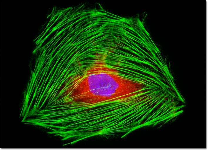
Male Rat Kangaroo Kidney Epithelial Cells (PtK2)
|
The dramatic advances in modern fluorophore technology are exemplified by the Alexa Fluor dyes. These sulfonated rhodamine derivatives exhibit higher quantum yields for more intense fluorescence emission than spectrally similar probes, and have several additional improved features, including enhanced photostability, absorption spectra matched to common laser lines, pH insensitivity, and a high degree of water solubility. In fact, the resistance to photobleaching of Alexa Fluor dyes is so dramatic that even when subjected to irradiation by high-intensity laser sources, fluorescence intensity remains stable for relatively long periods of time in the absence of antifade reagents. This feature enables the water soluble Alexa Fluor probes to be readily utilized for both live-cell and tissue section investigations, as well as in traditional fixed preparations. The single rat kangaroo kidney epithelial cell that appears in the digital image presented above was resident in a culture fluorescently labeled with Texas Red conjugated to the enzyme DNase I, targeting globular (unpolymerized) actin. The specimen was also labeled for filamentous actin with Alexa Fluor 488 conjugated to phalloidin, and for nuclear DNA with DAPI. Images were recorded in grayscale with a QImaging Retiga Fast-EXi camera system coupled to an Olympus BX-51 microscope equipped with bandpass emission fluorescence filter optical blocks provided by Omega Optical. During the processing stage, individual image channels were pseudocolored with RGB values corresponding to each of the fluorophore emission spectral profiles. |
© 1995-2025 by Michael W. Davidson and The Florida State University. All Rights Reserved. No images, graphics, software, scripts, or applets may be reproduced or used in any manner without permission from the copyright holders. Use of this website means you agree to all of the Legal Terms and Conditions set forth by the owners.
This website is maintained by our
|
