Fluorescence Digital Image Gallery
Normal Rat Kidney Epithelial Cells (NRK LIne)
|
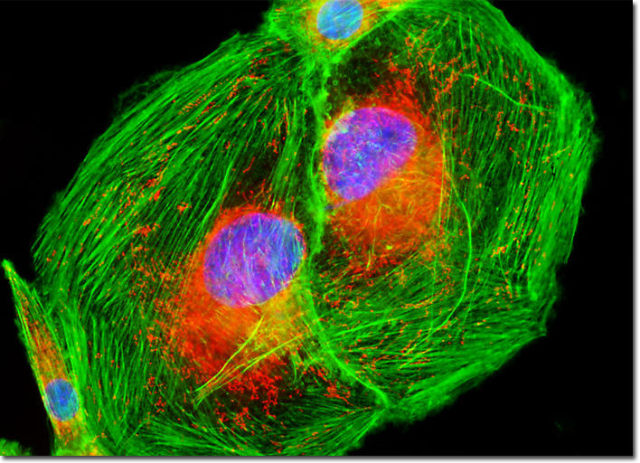
Normal kidney cells, such as those of the NRK line, are commonly utilized in laboratories around the world. They are extremely useful in the study of normal renal anatomy and physiology, as well as morphometric alterations related to age and the development of renal diseases, including renal dysplasia, pyelonephritis, azotemia, and diabetic nephropathy. Kidney cell lines have also been of significant interest due to scientific efforts to grow working kidneys in specimens in which the organs are absent or irreparably damaged. Such research endeavors may eventually lead to an abundant supply of kidneys for transplant purposes, eliminating the extremely long lists that are currently utilized to keep track of anxiously waiting patients. Each year thousands of people die waiting for a suitable kidney to become available. The rat kidney epithelial cells presented in the digital image above were resident in a culture stained with MitoTracker Red CMXRos, Alexa Fluor 488 conjugated to phalloidin, and DAPI, targeting mitochondria, the cytoskeletal filamentous actin network, and nuclear DNA, respectively. Images were recorded in grayscale with a QImaging Retiga Fast-EXi camera system coupled to an Olympus BX-51 microscope equipped with bandpass emission fluorescence filter optical blocks provided by Omega Optical. During the processing stage, individual image channels were pseudocolored with RGB values corresponding to each of the fluorophore emission spectral profiles. |
© 1995-2025 by Michael W. Davidson and The Florida State University. All Rights Reserved. No images, graphics, software, scripts, or applets may be reproduced or used in any manner without permission from the copyright holders. Use of this website means you agree to all of the Legal Terms and Conditions set forth by the owners.
This website is maintained by our
|
