Fluorescence Digital Image Gallery
Normal Rat Kidney Epithelial Cells (NRK LIne)
|
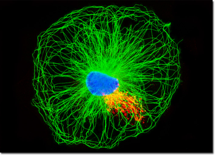
The human kidney is comprised of 12 or more sections called renal pyramids. A renal pyramid is a triangular sector of a kidney that comprises the medulla of the organ. The structure chiefly consists of numerous tubules that convey urine from the cortex of the kidney, where the waste product is generated, to cup-shaped cavities called calyces that hold and collect the urine until it eventually travels to the bladder via the ureter. The point of a renal pyramid, which contains numerous tiny openings that extend into a calyx, is known as a papilla. The small apertures allow urine to pass through the papilla into the calyx. Although human kidneys contain many renal pyramids, other animals may exhibit a significantly smaller number. For instance, the species Rattus norvegicus that is associated with the NRK cell line only features a single renal pyramid per kidney. The microtubules present in the culture of normal rat kidney cells featured in the digital image above were immunofluorescently labeled with primary anti-tubulin (pan) mouse monoclonal antibodies followed by goat anti-mouse Fab fragments conjugated to Alexa Fluor 488. Golgi bodies were simultaneously targeted with rabbit anti-giantin primary antibodies, followed by goat anti-rabbit secondaries conjugated to Texas Red. The ultraviolet-absorbing probe Hoechst 33342 was utilized to counterstain nuclei. Images were recorded in grayscale with a QImaging Retiga Fast-EXi camera system coupled to an Olympus BX-51 microscope equipped with bandpass emission fluorescence filter optical blocks provided by Omega Optical. During the processing stage, individual image channels were pseudocolored with RGB values corresponding to each of the fluorophore emission spectral profiles. |
© 1995-2025 by Michael W. Davidson and The Florida State University. All Rights Reserved. No images, graphics, software, scripts, or applets may be reproduced or used in any manner without permission from the copyright holders. Use of this website means you agree to all of the Legal Terms and Conditions set forth by the owners.
This website is maintained by our
|